
Drawing Of Ovaries
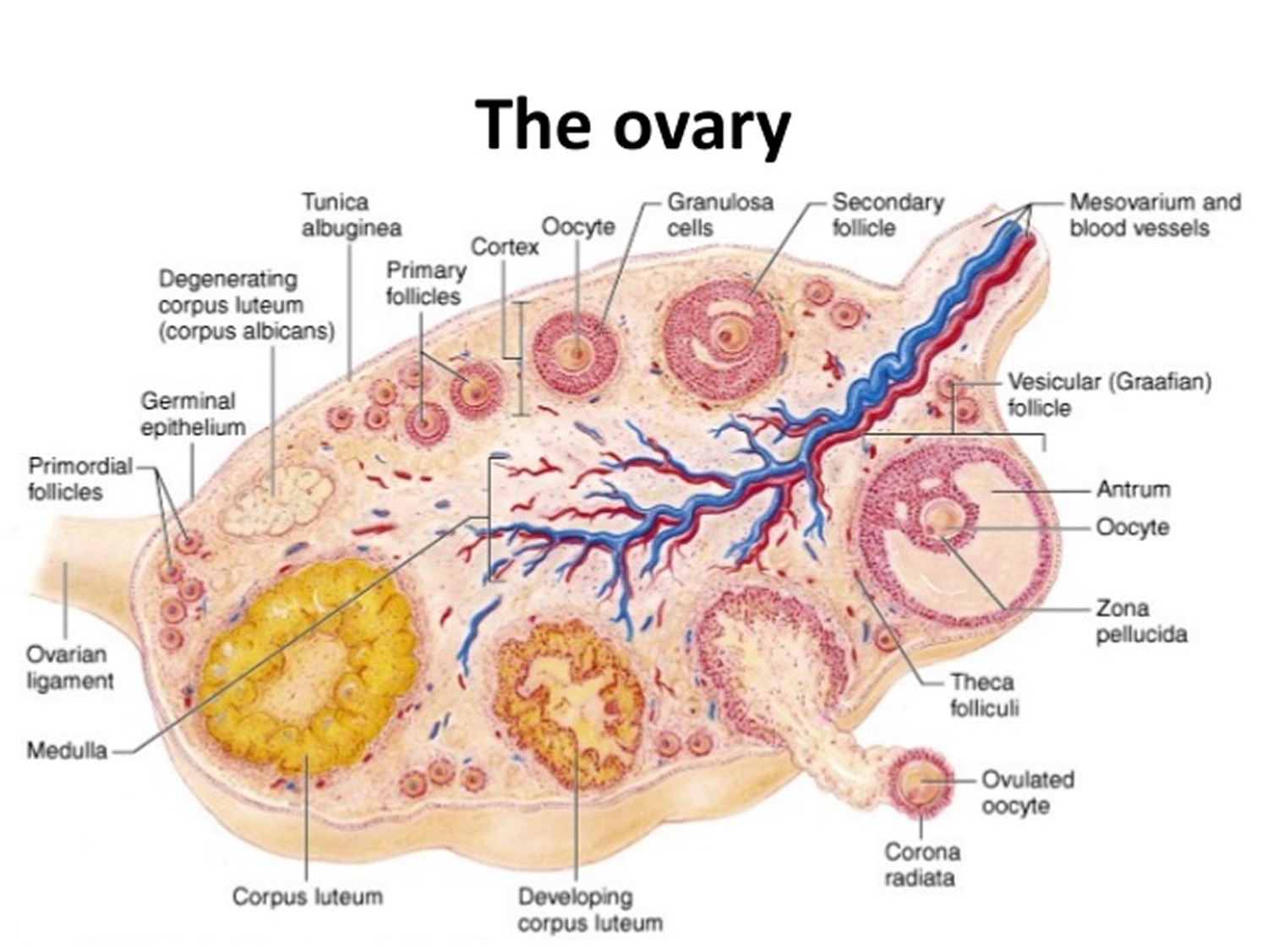
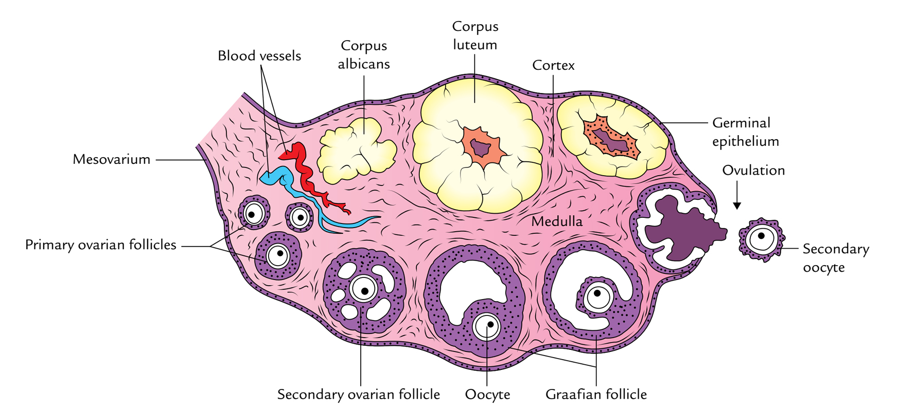
Drawing Of Ovaries - Inner to the tunica albuginea, the ovarian tissue is divided into two regions; Together they comprise the female reproductive system, supporting sexual and reproductive activities. Also, the ovaries act as an endocrine gland by releasing certain hormones in females. A centrally located medulla and a peripheral cortex. They play an important role in female hormone production and produce eggs during ovulation.
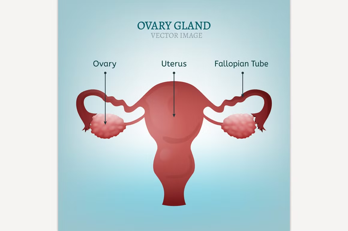
Web the ovaries are the female gonads (see figure 27.9). Most popular human anatomy female reproductive system, female reproductive. Web toggle anatomy system. The ovaries are located within the pelvic cavity, and are supported by the mesovarium, an extension of the peritoneum that connects the ovaries to the broad ligament. The ovaries produce eggs and also release hormones, such estrogen and progesterone. These organs are involved in the production and transportation of gametes and the production of sex hormones. Hairlike structures called cilia guide the egg from the ovary to the uterus.
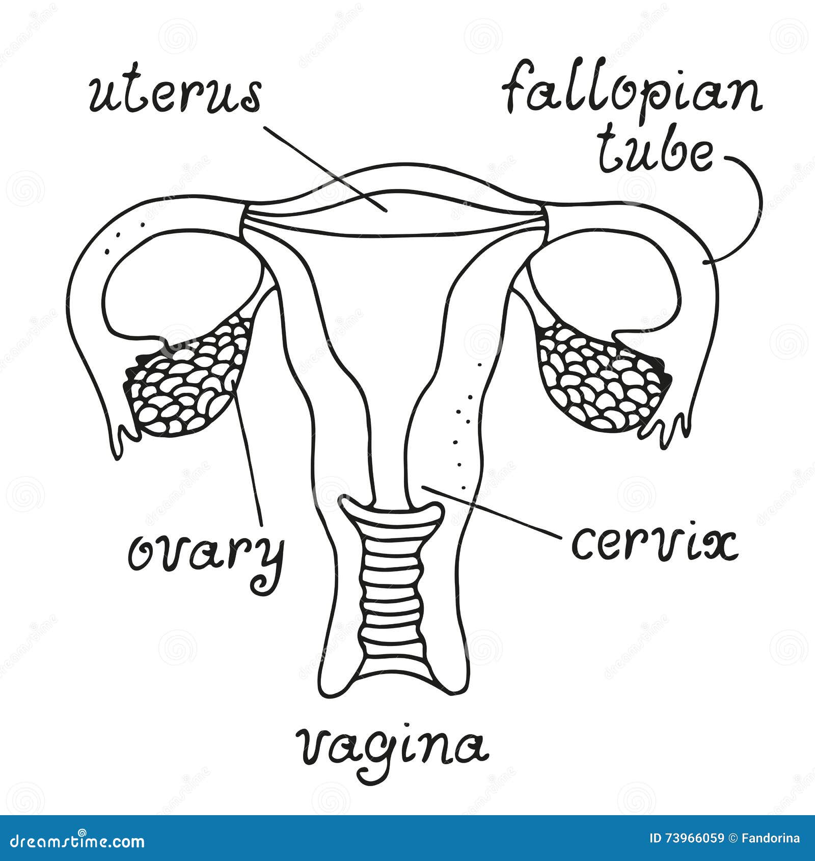
Uterus and ovaries anatomy stock vector. Illustration of medical 73966059
Your ovaries produce eggs and hormones. Hairlike structures called cilia guide the egg from the ovary to the uterus. Normal female reproductive system anatomy. Together they comprise the female reproductive system, supporting sexual and reproductive activities. The ovaries are located within the pelvic cavity, and are supported by the mesovarium, an extension of the peritoneum.
Ovary Gland Illustrations Creative Market
Web neet 2023 answer key ovary diagram the ovary is a primary gonad of the female reproductive system. #ovary #howtodraw #adimushowthis is an easy and simple drawing of a ovary. Also, the ovaries act as an endocrine gland by releasing certain hormones in females. The functions of these organs are involved in fertility, conception,. Using.

Schematic representation of the ovarian structure. Adapted from
Most popular human anatomy female reproductive system, female reproductive. They produce eggs and hormones for menstruation and pregnancy. The main functions of the ovaries are: They are attached on one edge, the hilus, to the broad ligament of the uterus by a fold of peritoneum, the mesovarium. The ovaries are small organs located on both.
Ovaries Function, Location, Hormones Produced. What control it?
Hairlike structures called cilia guide the egg from the ovary to the uterus. Paired ovals, they are each about 2 to 3 cm in length, about the size of an almond. The female reproductive system includes the ovaries, fallopian tubes, uterus, vagina, vulva, mammary glands and breasts. The ovum is fertilised by a motile sperm.

Ovary Diagrams to Print 101 Diagrams
Carry eggs from the ovaries to the uterus. Normal female reproductive system anatomy. Web vulva summary the female reproductive organs include several key structures, such as the ovaries, uterus, vagina, and vulva. Web the ovaries are the female gonads (see figure 27.9). None the female sex organs consist of both internal and external genitalia. The.

Uterus, Ovaries, Fallopian Tubes, Illustration Stock Image C043
Inner to the tunica albuginea, the ovarian tissue is divided into two regions; None the female sex organs consist of both internal and external genitalia. The fallopian tubes connect the ovaries to the uterus on each side. The female reproductive system also facilitates the fertilization of ova by. The main functions of the ovaries are:.
Ovaries Earth's Lab
Web the ovaries are the female gonads (see figure 27.9). Web the ovary is a ductless reproductive gland in which the female reproductive cells are produced. The female gonads (ovaries) are derivatives of the paramesonephric gonadal ridge. Web the ovaries are paired, oval organs attached to the posterior surface of the broad ligament of the.
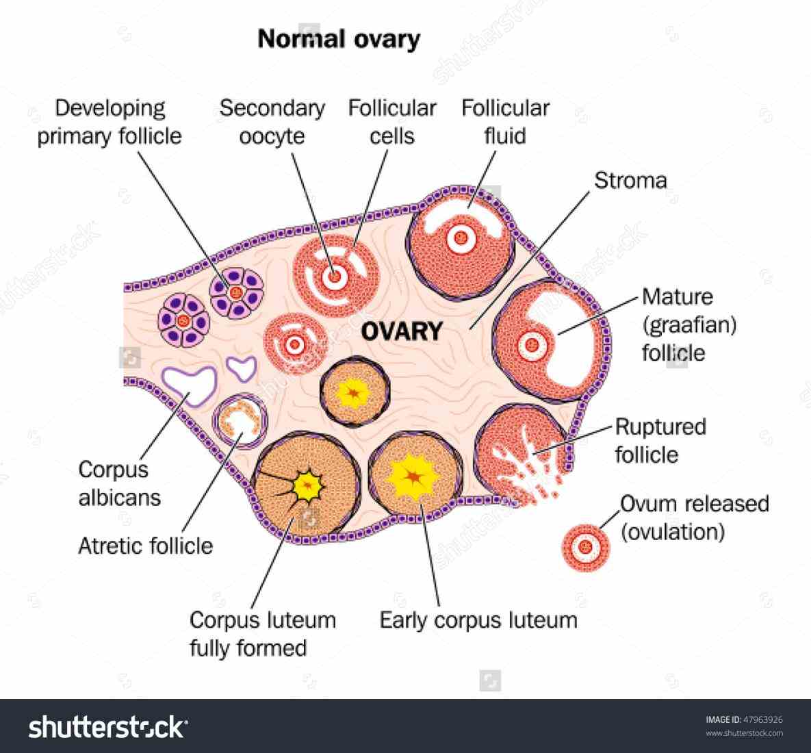
Draw A Labelled Diagram Of A Section Through Ovary
Web neet 2023 answer key ovary diagram the ovary is a primary gonad of the female reproductive system. It forms a canal that opens into the vagina, which leads to the outside of the body. You will also know the different characteristics of ovarian follicles in different animals. A centrally located medulla and a peripheral.
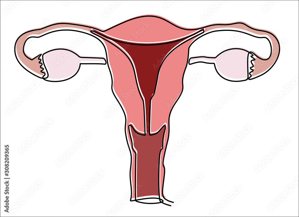
Continuous one single line drawing Uterus and ovaries, organs of female
The functions of these organs are involved in fertility, conception,. Web anatomy of the female reproductive system. Inner to the tunica albuginea, the ovarian tissue is divided into two regions; The lower, narrow part of the uterus (womb) located between the bladder and the rectum. Two female reproductive organs located in the pelvis. Web the.

Ovarian cycle. Yellow body corpus luteum (Drawing by W. Herzig
Paired ovals, they are each about 2 to 3 cm in length, about the size of an almond. Web it has three phases: The ovum is fertilised by a motile sperm to form a zygote. At which the ovarian follicle matures, and the hormone estrogen is secreted. Inner to the tunica albuginea, the ovarian tissue.
Drawing Of Ovaries Web overview the ovaries are small glands located on either side of your uterus. Using slide 239, examine the overall topography of the ovary and note the numerous vessels which enter it via the broad ligament. The uterus has a muscular outer layer called the myometrium and an inner lining called the endometrium. Inner to the tunica albuginea, the ovarian tissue is divided into two regions; Web in this single article you will get all the identifying characteristics of ovarian cortex, ovarian medulla and different follicles with real ovary histology slide pictures and labeled diagram.
Your Ovaries Produce Eggs And Hormones.
Most popular uterus female reproductive system. The ovaries are located within the pelvic cavity, and are supported by the mesovarium, an extension of the peritoneum that connects the ovaries to the broad ligament. Web the ovaries are the female gonads (see figure 27.9). They become distinct ovaries between the 6th and 7th gestational weeks, when epiblastic primordial germ.
Inner To The Tunica Albuginea, The Ovarian Tissue Is Divided Into Two Regions;
Web toggle anatomy system. Also, the ovaries act as an endocrine gland by releasing certain hormones in females. Neurovascular structures enter the hilum of the ovary via the mesovarium. The ovaries are paired organs situated on either side of the uterus.
Using Slide 239, Examine The Overall Topography Of The Ovary And Note The Numerous Vessels Which Enter It Via The Broad Ligament.
Carry eggs from the ovaries to the uterus. They produce eggs and hormones for menstruation and pregnancy. The female reproductive system includes the ovaries, fallopian tubes, uterus, vagina, vulva, mammary glands and breasts. Cup internal genitals of women
The Functions Of These Organs Are Involved In Fertility, Conception,.
None the female sex organs consist of both internal and external genitalia. Most popular human anatomy female reproductive system, female reproductive. The ovaries are small organs located on both sides of the pelvis. Organs location scheme uterus, cervix, ovary, fallopian tube.