
Femur Bone Drawing
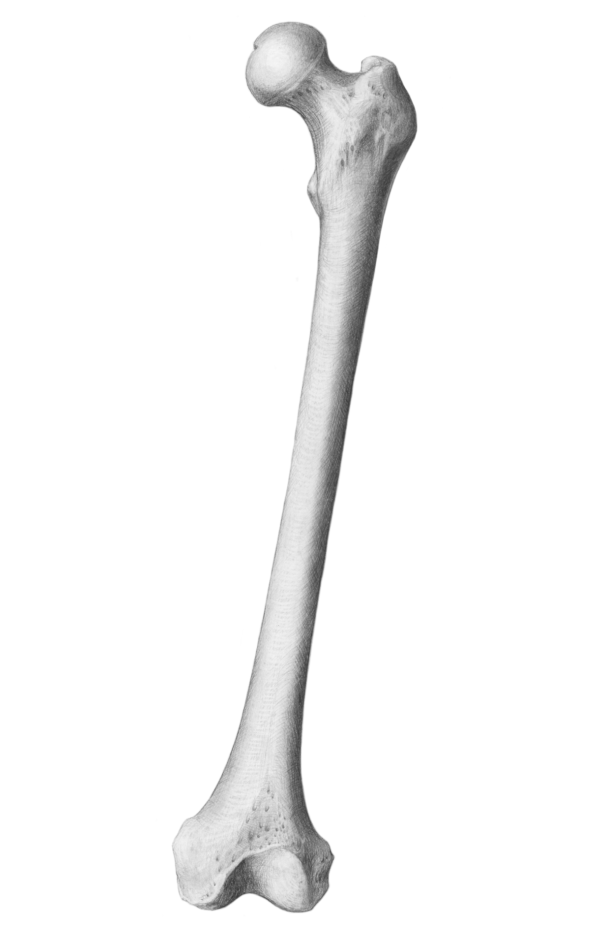
Femur Bone Drawing - I draw the femur bone with pencil on art paper. Let’s walk through the stick figure starting at the head (superior/proximal) and moving to the legs (inferior/distal). 1 waiting premieres may 20, 2023. The femur has two rounded ends and a long shaft in the middle. —the upper extremity presents for examination a head, a neck, a greater and a lesser trochanter.
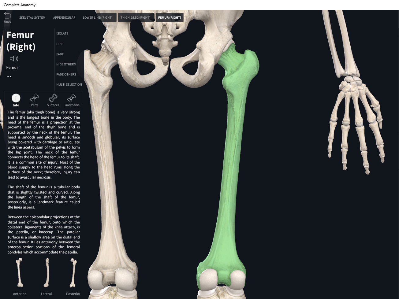
The femur is the only bone located within the human thigh. Web this angle of inclination ensures that the weight of the upper body passes along the mechanical axis of the femur. Web anatomy where is the femur located? Web the femur is the longest and strongest bone in the human body, extending from the hip to the knee. Drawing data is taken from ct scan in order to generate 3 d model of the femur bone structure using solid works. Learn how to draw the femur, patella, tibia, and fibula in this. Most popular antique illustration of human body anatomy bones:
Scientific Illustration jennifersmithart Bones Femur, Scapula, and...
Femur set of human leg bones set of human leg bones isolated on white knee joint icon Pelvis (female) human anatomy scientific illustrations with latin/italian labels: Below the head of the femur is the neck and the greater trochanter. I draw the femur bone with pencil on art paper. Femur set of human leg bones.

Femur (by lichtopdezaak) Learn and Remember
Most popular antique illustration of human body anatomy bones: Due to complex shape and structure of femur bone which consists of bone shaft, neck and tissues, we used finite element analysis (fea) to study biomechanical behaviour of femur bone. At the top, the femur has a rounded head that fits into the hip joint, allowing.
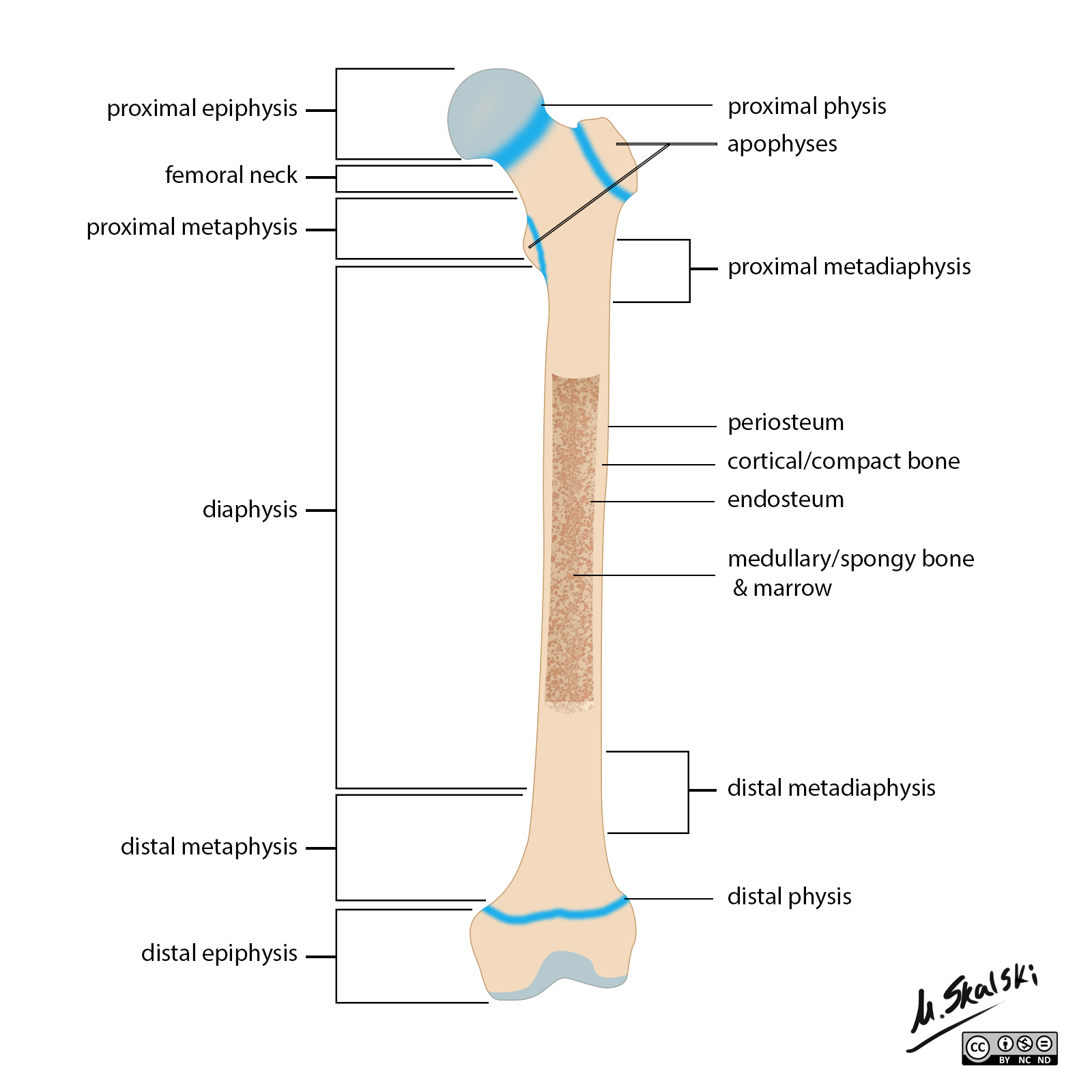
Radiopaedia Drawing Anatomy of long bones (femur) English labels
Web femur bone cut in half showing normal bone density and osteoporosis. The femur is the only bone in your thigh. When you break the femur bone at an angle. The femur is the only bone located within the human thigh. Web the femur is the only bone in the thigh and the longest bone.

Femur Bone Illustration Stock Illustration Download Image Now Femur
Click and start learning now! Due to complex shape and structure of femur bone which consists of bone shaft, neck and tissues, we used finite element analysis (fea) to study biomechanical behaviour of femur bone. It is both the longest and the strongest bone in the human body, extending from the hip to the knee..
Bones Femur. Anatomy & Physiology
The femur has two rounded ends and a long shaft in the middle. Web the femur, like other long bones, is divisible into a body and two extremities. Pelvis (female) human anatomy scientific illustrations with latin/italian labels: Click and start learning now! As you draw the femur, keep these features in mind: Femur set of.

2 Crosssection of a human proximal femur (left image) and scheme of
Web this angle of inclination ensures that the weight of the upper body passes along the mechanical axis of the femur. A cylinder with two round bumps at each end. I draw the femur bone with pencil on art paper. This axis can be identified by drawing a vertical line from the center of the.

Learn Femur diagram (by wawezase) Remember and Understand
Due to complex shape and structure of femur bone which consists of bone shaft, neck and tissues, we used finite element analysis (fea) to study biomechanical behaviour of femur bone. Femur set of human leg bones set of human leg bones isolated on white knee joint icon The femur is the only bone in your.

femur Definition, Function, Diagram, & Facts Britannica
Advertisement what does the femur look like? Web this angle of inclination ensures that the weight of the upper body passes along the mechanical axis of the femur. It’s the classic shape used for bones in cartoons: It acts as the site of origin and attachment of many muscles and ligaments, and can be divided.

Human femur bones, vector hand drawn illustration isolated on a white
The different parts of the human stick figure correlate with different parts of the femur. It’s the classic shape used for bones in cartoons: Femur set of human leg bones set of human leg bones isolated on white knee joint icon Web there is a simple way to remember the main anatomical features of the.
Biology Diagrams,Images,Pictures of Human anatomy and physiology Femur
Web an interactive tutorial featuring the anterior and posterior markings of the femur bone, with the aid of the iconic getbodysmart illustrations. Web anatomy of the femur, or thigh bone, made easy using a colored labeled diagram and drawing. There are also two prominent bony protrusions, the greater and lesser trochanter, that attach to muscles.
Femur Bone Drawing Ebraheim’s educational animated video describes anatomy and how to draw the femur.the lesser trochanter is a small rounded bump that is found on the post. Due to complex shape and structure of femur bone which consists of bone shaft, neck and tissues, we used finite element analysis (fea) to study biomechanical behaviour of femur bone. Web there is a simple way to remember the main anatomical features of the femur using a human stick figure as drawn below. 1 waiting premieres may 20, 2023. Web how to draw femur bone human step by step/femur bone drawing/human femur bone it is very easy drawing detailed method to help you.
Femur Broken In A Horizontal Line Directly Through The Shaft.
This axis can be identified by drawing a vertical line from the center of the femoral head to the center of a horizontal line across the tibial plateau (the center of the knee joint line). Due to complex shape and structure of femur bone which consists of bone shaft, neck and tissues, we used finite element analysis (fea) to study biomechanical behaviour of femur bone. Skeletal system anatomy of lower limb for nursing, medical learners, usmle, and more! Learn how to draw the femur, patella, tibia, and fibula in this.
Web Browse 280+ Human Femur Bone Drawing Stock Photos And Images Available, Or Start A New Search To Explore More Stock Photos And Images.
It acts as the site of origin and attachment of many muscles and ligaments, and can be divided into three parts; As you draw the femur, keep these features in mind: Web femur bone cut in half showing normal bone density and osteoporosis. Ebraheim’s educational animated video describes anatomy and how to draw the femur.the lesser trochanter is a small rounded bump that is found on the post.
Web Common Types Of Femur Fractures:
Most popular antique illustration of human body anatomy bones: Drawing data is taken from ct scan in order to generate 3 d model of the femur bone structure using solid works. How to draw femur bone human/femour bone human bodyit is very easy drawing detailed method to help you.i draw the femur bone with pencil on art paper on my e. A cylinder with two round bumps at each end.
I Draw The Femur Bone With Pencil On Art Paper.
Femur set of human leg bones set of human leg bones isolated on white knee joint icon Web the femur is the only bone in the thigh and the longest bone in the body. Web anatomy of the femur, or thigh bone, made easy using a colored labeled diagram and drawing. It runs from your hip to your knee.