
Foot Bones Drawing
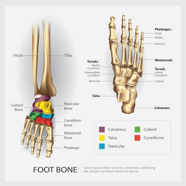
Foot Bones Drawing - Select from premium foot bone diagram images of the highest quality. Web they can be divided into three groups: The intermediate tarsal bone is the navicular. Web fortunately, the bones are a great way to study the foot. The hindfoot is the back portion of the foot, and it includes the heel, which connects the foot to the lower leg.
The intermediate tarsal bone is the navicular. Learn more about foot bones and foot anatomy here. The proximal tarsal bones are the talus and calcaneus. Free for commercial use high quality images. How to draw feet basics of the foot. Web fortunately, the bones are a great way to study the foot. Web it’s time to learn how to draw a foot!

artist anatomy foot Iskanje Google Anatomy art, Skeleton drawings
The intermediate tarsal bone is the navicular. Find foot bone diagram stock illustrations from getty images. The bone structure of the foot consists of three different sections: Science & technology 3d models. Web all 26 bones of the foot are described generally for drawing purposes. The hindfoot, the midfoot, and the forefoot. A simple way.
Skeleton Feet Drawing at Explore collection of
Web it’s time to learn how to draw a foot! Find foot bone diagram stock illustrations from getty images. No toes, no arches, just the. Web human anatomy fundamentals: Web the feet support the human body when standing, walking, running, and more. Web vector illustration of bare male and female legs on white background. Web.

Skeleton Foot by Isasan on DeviantArt
The hindfoot, the midfoot, and the forefoot. Most popular bones of the foot and ankle joint medical vector illustration. Web it’s time to learn how to draw a foot! Web your assignment is to simplify the foot bones into their basic forms. Most popular bones of the foot and ankle joint medical vector illustration. Science.

Bones of the Foot Anatomy Sketch
These make up the toes and broad section of the feet. Select from premium foot bone diagram images of the highest quality. Most popular bones of the foot and ankle joint medical vector illustration. Since the foot is so bony, knowing the inside anatomy. The tarsals or ankle bones in blue, the metatarsi. A simple.

Foot Bone Anatomy Vector Illustration 539973 Vector Art at Vecteezy
Web all 26 bones of the foot are described generally for drawing purposes. The proximal tarsal bones are the talus and calcaneus. The bone structure of the foot consists of three different sections: Most popular human foot bones front and side view anatomy Web vector illustration of bare male and female legs on white background..
Foot Skeleton Drawing at GetDrawings Free download
No toes, no arches, just the. Most popular bones of the foot and ankle joint medical vector illustration. Characters & creatures 3d models. Web all 26 bones of the foot are described generally for drawing purposes. They are situated proximally in the foot in the ankle area. In this last body part of the anatomy.
.jpg)
Foot Bone Diagram resource Imageshare
Learn more about foot bones and foot anatomy here. Since the foot is so bony, knowing the inside anatomy. Web learn the bones of the foot in half the time with these interactive quizzes and labeling activities! Draw from life using your own feet or draw from the 3d models i provide you. Most popular.
Premium Vector Foot bone anatomy vector illustration
No toes, no arches, just the. Human foot bone foot bone structure foot bone diagram foot bone vector sort by: The hindfoot, the midfoot, and the forefoot. Select from premium foot bone diagram images of the highest quality. Since the foot is so bony, knowing the inside anatomy directly helps you draw the outside surface..
Bones of the Feet ClipArt ETC
The intermediate tarsal bone is the navicular. Since the foot is so bony, knowing the inside anatomy. How to draw feet basics of the foot. The bone structure of the foot consists of three different sections: The skeletal structure of the foot. Web all 26 bones of the foot are described generally for drawing purposes..
.jpg)
Foot Bone Diagram resource Imageshare
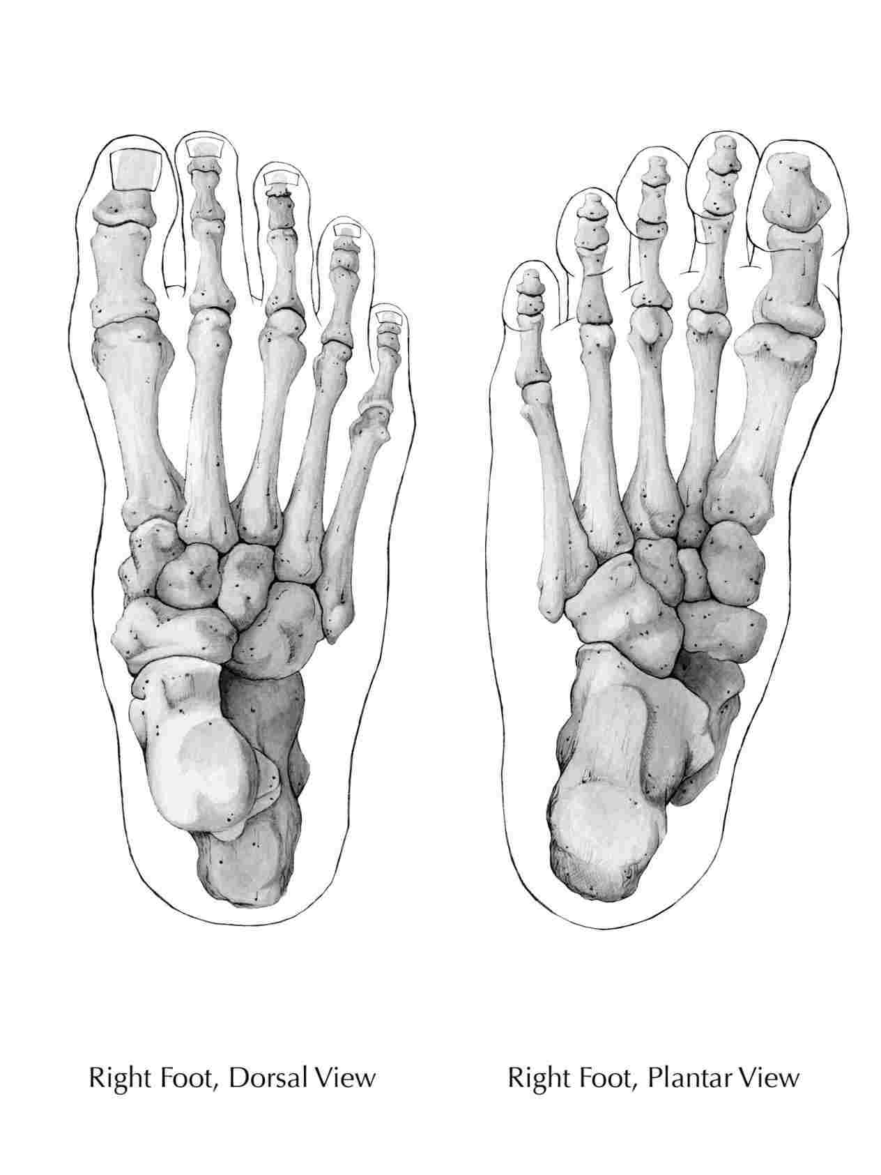
Human foot bone foot bone structure foot bone diagram foot bone vector sort by: Web human anatomy fundamentals: Web the 26 bones of the foot consist of eight distinct types, including the tarsals, metatarsals, phalanges, cuneiforms, talus, navicular, and cuboid bones. Free for commercial use high quality images. Web the foot is challenging to draw.
Foot Bones Drawing The proximal tarsal bones are the talus and calcaneus. Science & technology 3d models. Draw from life using your own feet or draw from the 3d models i provide you. Web a medical illustration of a healthy foot and a foot affected by heel bursitis. Web it’s time to learn how to draw a foot!
Web All 26 Bones Of The Foot Are Described Generally For Drawing Purposes.
This lesson will focus on the overall design of the foot, and the form, proportion, and mobility of the individual bones. The hindfoot, the midfoot, and the forefoot. These make up the toes and broad section of the feet. The intermediate tarsal bone is the navicular.
Since The Foot Is So Bony, Knowing The Inside Anatomy.
Web a medical illustration of a healthy foot and a foot affected by heel bursitis. No toes, no arches, just the. Web the 26 bones of the foot consist of eight distinct types, including the tarsals, metatarsals, phalanges, cuneiforms, talus, navicular, and cuboid bones. Web vector illustration of bare male and female legs on white background.
Realistic Bones Of Foot Skeleton Of Human Leg.
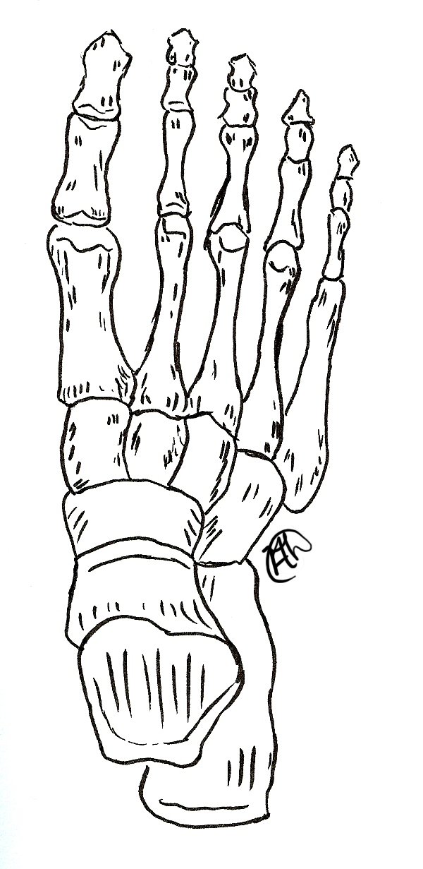
Web browse 540+ foot bones drawing stock photos and images available, or start a new search to explore more stock photos and images. In this last body part of the anatomy course you’ll learn how to construct the foot with basic forms, l. Let’s look briefly at the structure of the foot: Human foot bone foot bone structure foot bone diagram foot bone vector sort by:
The Hindfoot Is The Back Portion Of The Foot, And It Includes The Heel, Which Connects The Foot To The Lower Leg.
The bone structure of the foot consists of three different sections: The bones of the foot. Bones of the foot and ankle joint medical vector illustration isolated on white background eps 10 human skeleton structure. Web learn the bones of the foot in half the time with these interactive quizzes and labeling activities!