
Heart Drawing Biology
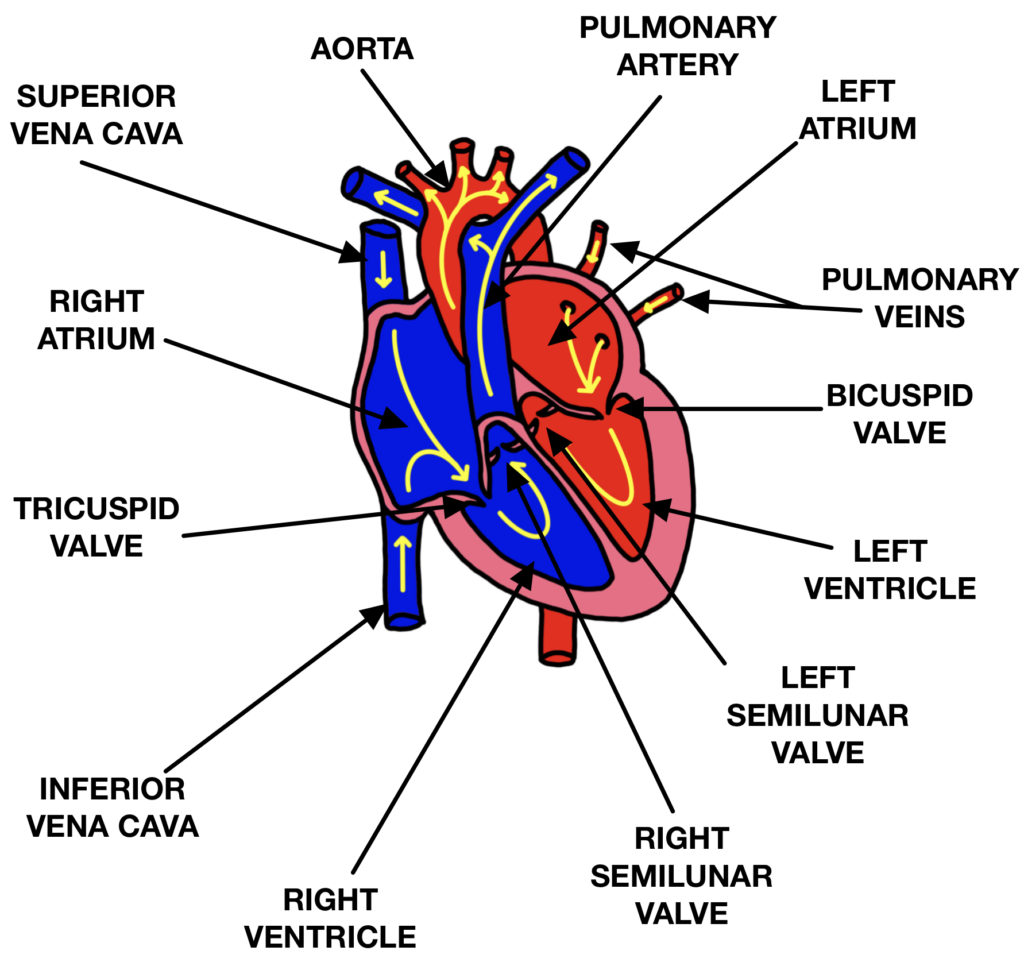
Heart Drawing Biology - Angle the slightly tampered end of the shape to the left about 120 degrees. The middle layer of the heart wall is called myocardium. These arcs will represent the chambers of the heart. The atria are the two superior chambers of the heart and the ventricles are the two inferior chambers of the heart. Valves are present to prevent the backflow of blood.
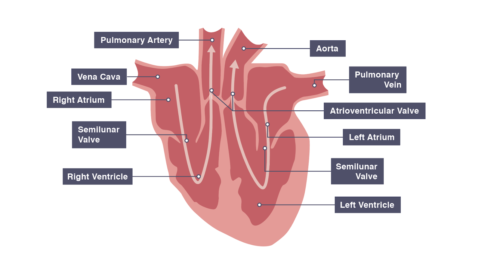
Students who are unwilling to handle hearts, but keen to see what is going on could take digital photographs of the. Web substances that alter blood pressure | chemicals, hormones, and neurotransmitters. Web the heart has four chambers, and most diagrams will show the heart as it is viewed from the ventral side. Use a pen or pencil to draw the heart's main body. The heart is divided into four chambers. Web the human heart has four chambers and is separated into two halves by the septum. Web the structure of the heart if you clench your hand into a fist, this is approximately the same size as your heart.

The human heart Biology assignment YouTube
Web 1.2k views 7 days ago. Draw two smaller arcs on the left and right sides of the circle. The middle layer of the heart wall is called myocardium. Whether you’re an aspiring artist or a medical student, understanding the anatomy and function of the heart is essential. Web prepare a handout with a drawing.
Heart Contractions Simplified Interactive Biology, with Leslie Samuel
This circle will represent the heart’s outline. Web objective while drawing a heart, learn the basic parts of the heart and how blood circulates throughout the body. Draw two smaller arcs on the left and right sides of the circle. These arcs will represent the chambers of the heart. Hi everyone, in this video i.
DRAW IT NEAT How to draw human heart labeled
These arcs will represent the chambers of the heart. Web about press copyright contact us creators advertise developers terms privacy policy & safety how youtube works test new features nfl sunday ticket press copyright. The atria are the two superior chambers of the heart and the ventricles are the two inferior chambers of the heart..

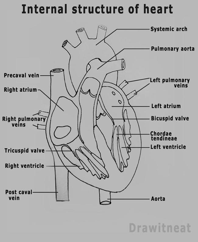
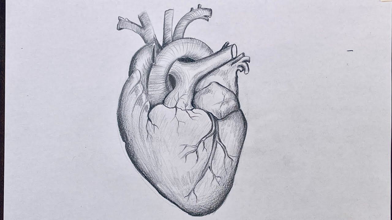
How to Draw the Internal Structure of the Heart 14 Steps
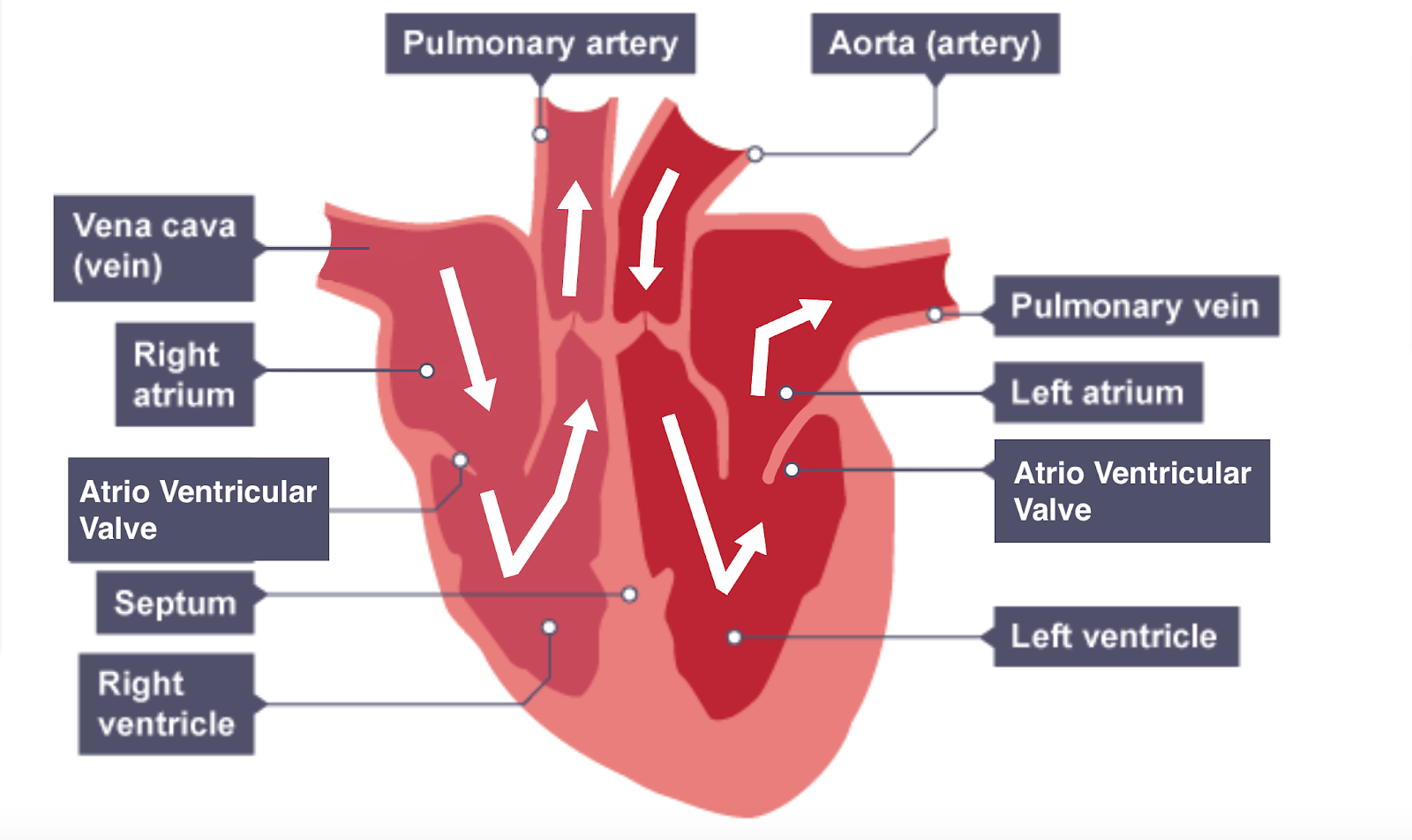
The left and right sides of the heart are separated by a wall of muscular tissue, called the septum. Web the heart is a unidirectional pump. Learning how to draw a human heart can be a challenging but rewarding experience. In this activity, your students will learn the basic anatomy of the heart and how.
IGCSE Biology 2017 2.65 Describe the Structure of the Heart and How
The outer layer of the heart wall is called epicardium. ©grey’s digital online, llc for more great resources visit: Web the heart is made up of four chambers: Web the heart drawing activity. The right side pumps deoxygenated blood (low in oxygen and high in carbon dioxide) to the lungs. These arcs will represent the.

human heart anatomy biology healthy image Stock Vector Image & Art
Web the heart is a unidirectional pump. Follow my step by step drawing tutorial and make your own xxxx drawing easy. Learning how to draw an anatomical heart will refine your drawing skills as you learn how to draw various complex components that the heart is made up of. It is located in the middle.

heart PMG Biology
The outer layer of the heart wall is called epicardium. The upper two chambers of the heart are called auricles. Your drawing will be simplified so you do not need to be an artist to do this! Next, they will add the four chambers of the heart. Whether you’re an aspiring artist or a medical.
IGCSE Biology Notes 2.63 Describe the Structure of the Heart and How
Here we will review its essential components, and how and why blood passes through them. Web the human heart has four chambers and is separated into two halves by the septum. Web the heart has four chambers, and most diagrams will show the heart as it is viewed from the ventral side. We can see.
Structure of the Heart The Science and Maths Zone
Your drawing will be simplified so you do not need to be an artist to do this! This is a quick way to learn how to draw the heart and some of the associated structures. Web about press copyright contact us creators advertise developers terms privacy policy & safety how youtube works test new features.
How to draw Heart Biology drawing for science students YouTube
The heart and the circulatory system together form the cardiovascular system. Whether you’re an aspiring artist or a medical student, understanding the anatomy and function of the heart is essential. Web objective while drawing a heart, learn the basic parts of the heart and how blood circulates throughout the body. We can see the heart.
Heart Drawing Biology Learn more about the heart in this article. The upper two chambers of the heart are called auricles. No doubt, the heart is the most important organ in our body. Students will start the activity by drawing a typical valentine’s day style heart. Web the heart is divided into four chambers:
The Two Top Chambers Are Atria And The Bottom Two Chambers Are Ventricles.
Web the structure of the heart if you clench your hand into a fist, this is approximately the same size as your heart. The heart wall is made up of three layers: No doubt, the heart is the most important organ in our body. Arrange your paper landscape like this!
Angle The Slightly Tampered End Of The Shape To The Left About 120 Degrees.
11m views 8 years ago. The left and right sides of the heart are separated by a wall of muscular tissue, called the septum. This circle will represent the heart’s outline. Web the heart’s unique design allows it to accomplish the incredible task of circulating blood through the human body.
In This Lecture, Dr Mike Shows The Two Best Ways To Draw And Label The Heart!
Learning how to draw a human heart can be a challenging but rewarding experience. Next, they will add the four chambers of the heart. Here we will review its essential components, and how and why blood passes through them. The heart is divided into four chambers.
Web About Press Copyright Contact Us Creators Advertise Developers Terms Privacy Policy & Safety How Youtube Works Test New Features Nfl Sunday Ticket Press Copyright.
The outer layer of the heart wall is called epicardium. **for each of the numbers described below, label on the heart diagram.**. The atria are the two superior chambers of the heart and the ventricles are the two inferior chambers of the heart. Layers of the heart wall.