
Hyaline Cartilage Drawing
Hyaline Cartilage Drawing - This article will focus on important features of hyaline cartilage, namely its matrix, chondrocytes, and perichondrium. 5.4k views 3 years ago. Web now, i will provide you with the drawing tutorial of hyaline cartilage slide images. Web during embryonic development, hyaline cartilage serves as temporary cartilage models that are essential precursors to the formation of most of the axial and appendicular skeleton. 20 format_list_bulleted contents add cartilage is flexible connective tissue found throughout the whole body.
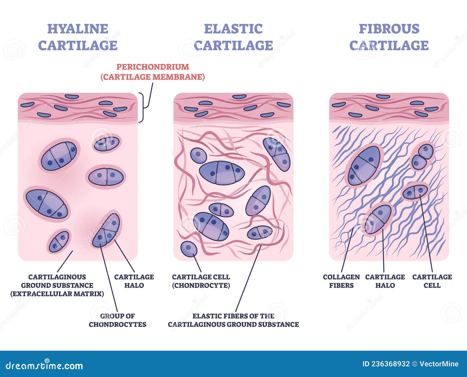
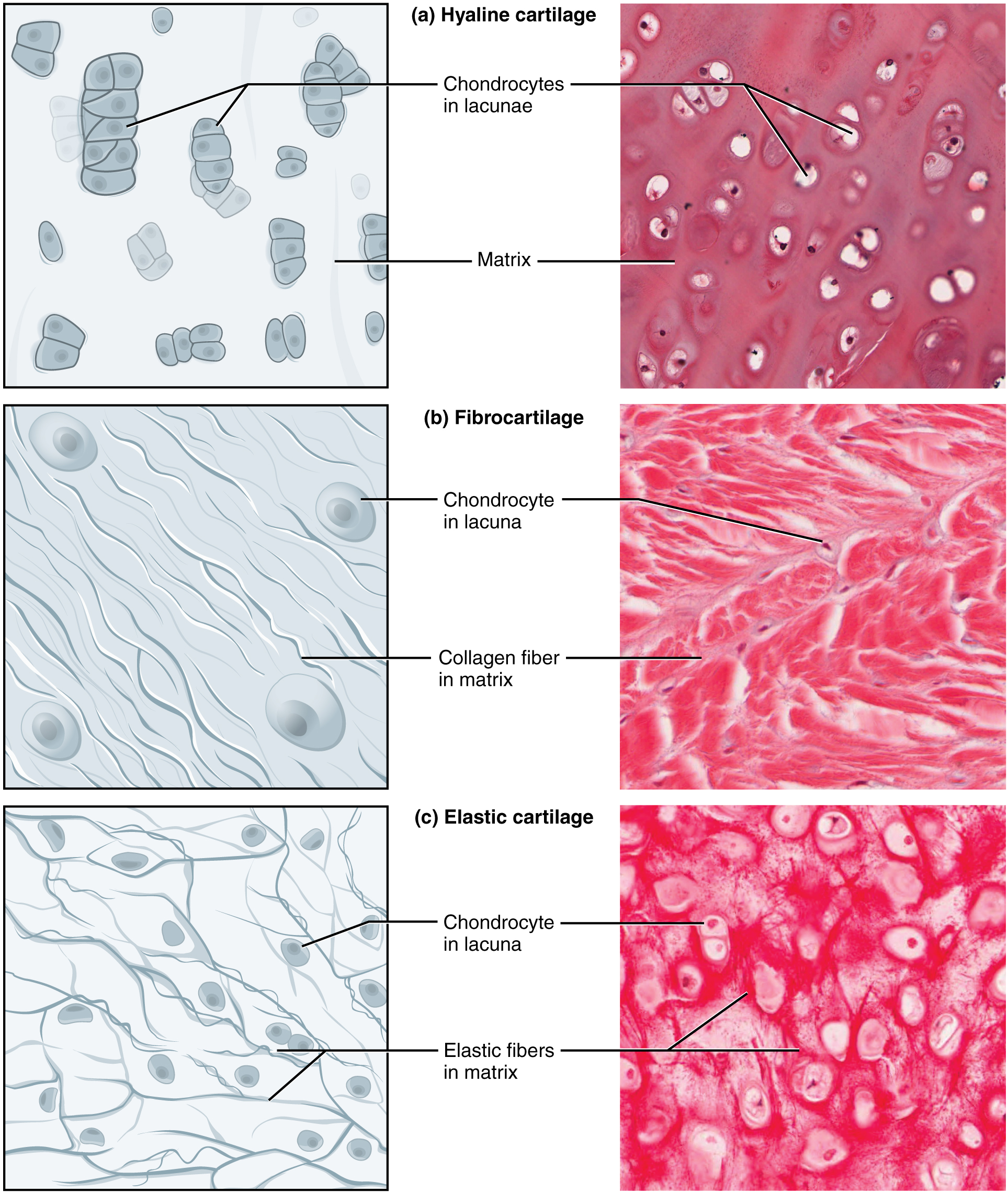
The other two types are elastic cartilage and fibrocartilage. Hyaline cartilage stock photos are available in a variety of. Web likecomment share subscribe #hyalinecartilage #histodiagrams #hyalinecartilagediagram #cartilagehistology Web obtain a slide of hyaline cartilage connective tissue from the slide box. This image shows a cross section of a cartilage ring that supports the trachea and maintains the openness (patency) of. Web publish with us. Hyaline cartilage is a supportive connective tissue with a rigid yet slightly flexible extracellular matrix.

Schematic drawing of articular (hyaline) cartilage containing abundant
This image shows a cross section of a cartilage ring that supports the trachea and maintains the openness (patency) of. Ac is a dense connective tissue mainly comprised of collagen, proteoglycans, organized in special zones containing special types of cells called articular chondrocytes [1,2].the biological and mechanical properties. Territorial matrix lies immediately around each isogenous.

Hyaline Cartilage Drawing YouTube
Fill out the blanks next to your drawing. Web introduction to hyaline cartilage. Hyaline cartilage stock photos are available in a variety of. The cells are called chondrocytes (ch) and the spaces in the cartilage in which they are found are called lacunae. Isogenous groups and interstitial growth results when chondrocytes divide and produce extracellular.
Cartilage types a)Hyaline Cartilage Cartilage System
It is also most commonly found in the ribs, nose, larynx, and trachea. Web elastic cartilage histology labeled diagram and drawing hyaline cartilage structure function of cartilage conclusion elastic cartilage histology first, you should know the important structures of elastic cartilage that. This image shows a cross section of a cartilage ring that supports the.
Perichondrium As Hyaline and Elastic Cartilage Membrane Outline Diagram
5.4k views 3 years ago. Its principal function is to provide a smooth, lubricated surface. Web introduction to hyaline cartilage. The cells are called chondrocytes (ch) and the spaces in the cartilage in which they are found are called lacunae. It contains no nerves or blood vessels, and its structure is relatively simple. Web hyaline.

Hyaline Cartilage, Vintage Illustration Stock Vector Illustration of
Hyaline cartilage is a supportive connective tissue with a rigid yet slightly flexible extracellular matrix. A joint of the jaw that connects it to the temporal bones of the skull. It is one of the three types of cartilage; Web introduction to hyaline cartilage. The cells are called chondrocytes (ch) and the spaces in the.
Connective Tissue Supports and Protects · Anatomy and Physiology
Articular cartilage (ac) is a loadbearing soft tissue that overlies the interacting bony surfaces in diarthrodial joints. Web publish with us. Web introduction to hyaline cartilage. Web obtain a slide of hyaline cartilage connective tissue from the slide box. If you want, you may follow this simple cartilage drawing tutorial. The cells are called chondrocytes.

Hyaline cartilage structure and biochemical composition. Schematic
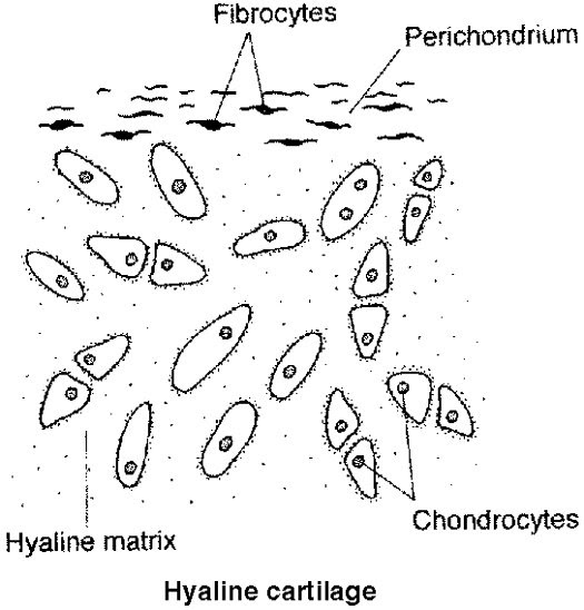
Web hyaline cartilage a higher magnification of the wall of the trachea shows the lumen with its epithelial lining in the lower left of the image. This article will focus on important features of hyaline cartilage, namely its matrix, chondrocytes, and perichondrium. In the circle below, draw a representative sample of key features you identified,.

Histology Image Cartilage
In the circle below, draw a representative sample of key features you identified, taking care to correctly and clearly draw their true shapes and directions. Cartilage grows by formation of additional matrix and incorporation of new cells from the inner chondrogenic layer of the perichondrium. Hyaline cartilage has very few fibers in. A type of.

How to Draw Hyaline Cartilage Simple and easy steps Biology Exam
Its principal function is to provide a smooth, lubricated surface. Ac is a dense connective tissue mainly comprised of collagen, proteoglycans, organized in special zones containing special types of cells called articular chondrocytes [1,2].the biological and mechanical properties. Hyaline cartilage is the most prevalent type, forming articular cartilages and the framework for parts of the.

Illustrations Hyaline Cartilage General Histology
Web hyaline cartilage has a smooth surface and is the most common of the three types of cartilage. Web hyaline cartilage, the most abundant type of cartilage, plays a supportive role and assists in movement. Hyaline cartilage is a supportive connective tissue with a rigid yet slightly flexible extracellular matrix. You might also read other.
Hyaline Cartilage Drawing Ac is a dense connective tissue mainly comprised of collagen, proteoglycans, organized in special zones containing special types of cells called articular chondrocytes [1,2].the biological and mechanical properties. Step by step drawing of histology of. 5.4k views 3 years ago. Hyaline cartilage is the most prevalent type, forming articular cartilages and the framework for parts of the nose, larynx, and trachea. Web hyaline cartilage a higher magnification of the wall of the trachea shows the lumen with its epithelial lining in the lower left of the image.
The Other Two Types Are Elastic Cartilage And Fibrocartilage.
It contains no nerves or blood vessels, and its structure is relatively simple. A joint of the jaw that connects it to the temporal bones of the skull. Hyaline cartilage stock photos are available in a variety of. Web hyaline cartilage, the most abundant type of cartilage, plays a supportive role and assists in movement.
It Is One Of The Three Types Of Cartilage;
A type of cartilage found on many joint surfaces; In the circle below, draw a representative sample of key features you identified, taking care to correctly and clearly draw their true shapes and directions. It is also most commonly found in the ribs, nose, larynx, and trachea. Web now, i will provide you with the drawing tutorial of hyaline cartilage slide images.
5.4K Views 3 Years Ago.
Web hyaline cartilage has a smooth surface and is the most common of the three types of cartilage. Articular cartilage (ac) is a loadbearing soft tissue that overlies the interacting bony surfaces in diarthrodial joints. You might also read other articles like elastic cartilage histology and fibrocartilage histology from the anatomy leaner blog. If you want, you may follow this simple cartilage drawing tutorial.
The Cells Are Called Chondrocytes (Ch) And The Spaces In The Cartilage In Which They Are Found Are Called Lacunae.
Formed by the process of chondrogenesis, the resulting chondrocytes are capable of producing large amounts of collagenous extracellular matrix and ground substance, which together form cartilage itself. Web cartilage consists of cells embedded in a matrix (mat) of fibers and ground substance. Web during embryonic development, hyaline cartilage serves as temporary cartilage models that are essential precursors to the formation of most of the axial and appendicular skeleton. Web elastic cartilage histology labeled diagram and drawing hyaline cartilage structure function of cartilage conclusion elastic cartilage histology first, you should know the important structures of elastic cartilage that.