Simple Columnar Epithelium Drawing
Simple Columnar Epithelium Drawing - These cells are found in the cornea, inner ear, and nose. Web simple columnar epithelial cells can specialize to secret mucus that coats and protects the surrounding surface from damage (insert link). Web there are two functionally different types of simple columnar epithelium: In humans, simple columnar epithelium lines most organs of the digestive tract including the stomach, and intestines. Because the epithelium can be innervated, simple columnar epithelium is also specialized to provide sensory input.
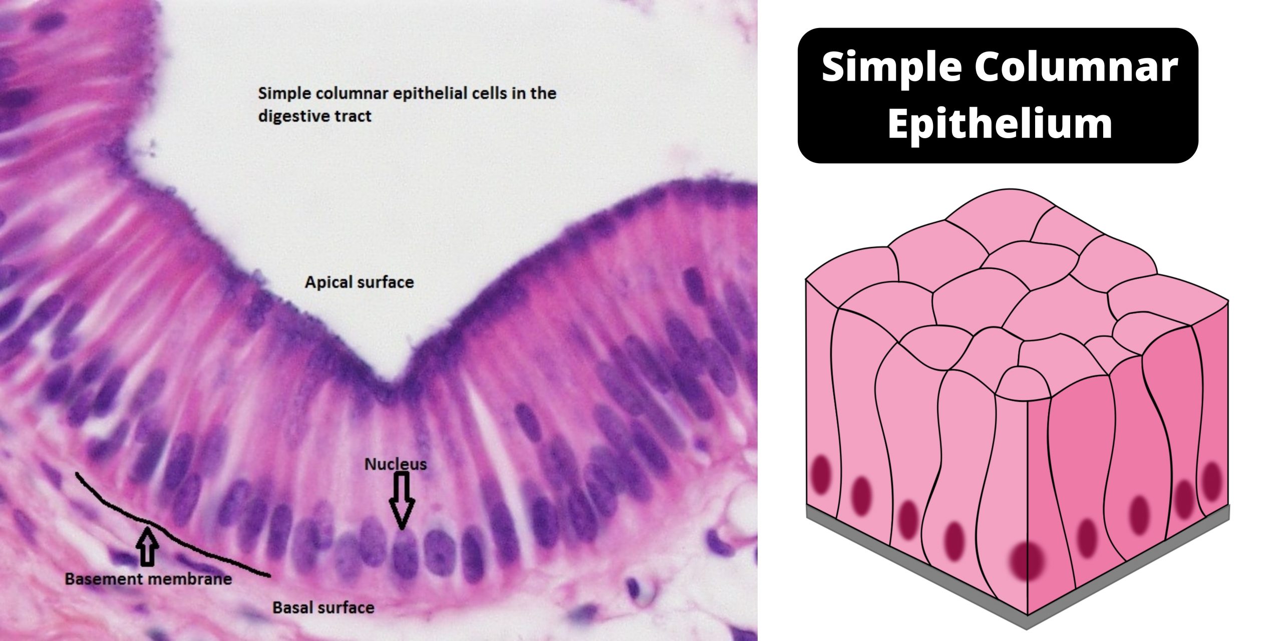
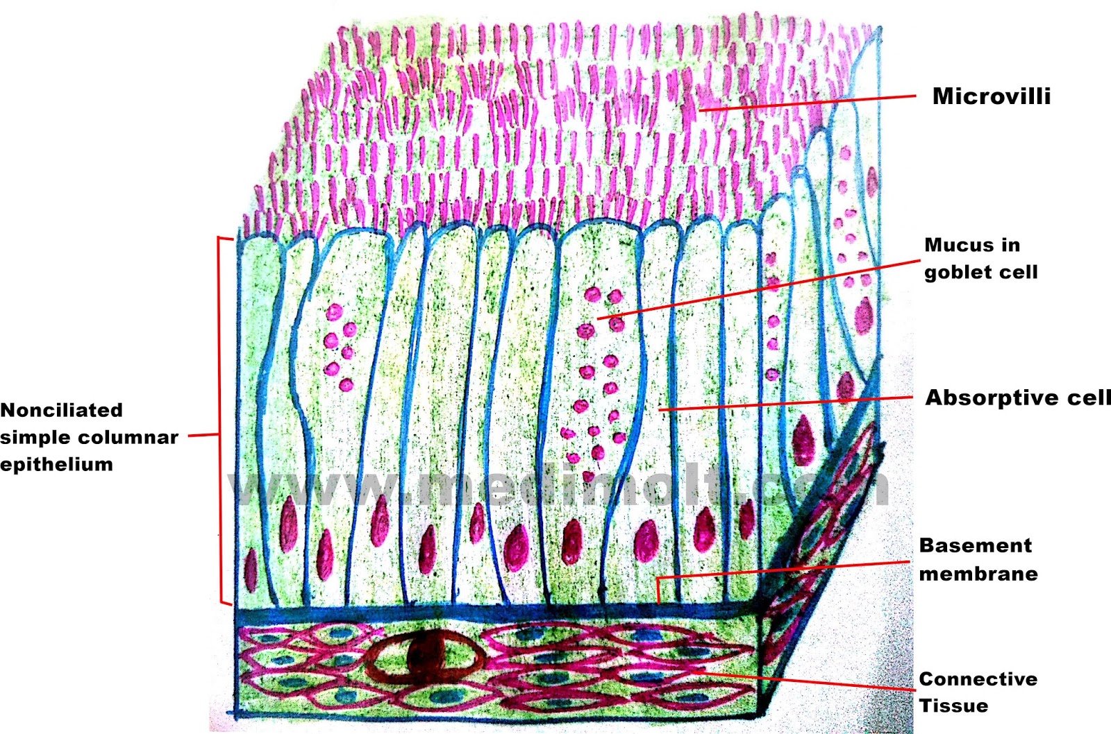
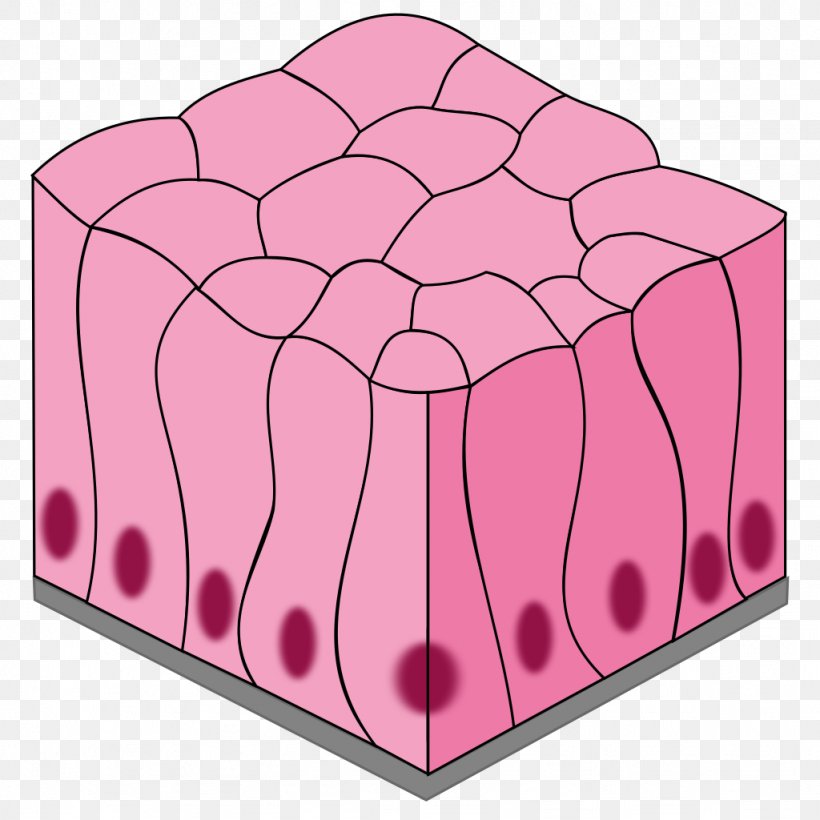
The cell remain deep but do not re. Web simple epithelium is one of the types of epithelium that is divided into simple columnar epithelium, simple squamous epithelium, and simple cuboidal epithelium. Like cuboidal epithelium, the cells in the columnar epithelium are also modified to suit the function and structure of the organ better. Like the cuboidal epithelia, this epithelium is active in the absorption and secretion of molecules. The entire layer of simple columnar epithelium is indicated by the bar. Web simple columnar epithelium (400x) primate small intestine the arrows are pointing to goblet cells that produce and release mucus. These are the single layer of cells with a greater height than breadth (columnar shape) and an oval basal nucleus.
Simple Columnar Epithelium (LM) Stock Image C022/2221 Science
Trachea and most of the upper respiratory tract (ciliated cells) Use the hotspot image below to learn more about the characteristics of simple columnar epithelium in the small intestine. Bodytomy provides a labeled diagram to help you understand the structure and function of simple columnar epithelium. The entire layer of simple columnar epithelium is indicated.
Simple columnar epithelium definition, structure, functions, examples
Typically, the simple columnar epithelium locates in the lining of the gastrointestinal tract, gall bladder, respiratory tract, uterine tube, and auditory tube. Web distinguish between simple epithelia and stratified epithelia, as well as between squamous, cuboidal, and columnar epithelia describe the structure and function of endocrine and exocrine glands Web a squamous epithelial cell looks.
Simple columnar epithelium, light micrograph Stock Image C052/8835
They look clear because the molecules in the mucus do not absorb a lot of stain. L=lumen n=nucleus read more pseudostratified columnar ciliated. Web simple columnar epithelium (400x) primate small intestine the arrows are pointing to goblet cells that produce and release mucus. These cells are found in the cornea, inner ear, and nose. Like.
Simple columnar epithelium Pseudostratified columnar epithelium Simple
Web simple columnar epithelium (400x) primate small intestine the arrows are pointing to goblet cells that produce and release mucus. Web simple columnar epithelial cells can specialize to secret mucus that coats and protects the surrounding surface from damage (insert link). A few epithelial layers are constructed from cells that are said to have a.
Simple Columnar Epithelium Description Single layer of elongated
Trachea and most of the upper respiratory tract (ciliated cells) Web simple columnar epithelia are tissues made of a single layer of long epithelial cells that are often seen in regions where absorption and secretion are important features. Like the cuboidal epithelia, this epithelium is active in the absorption and secretion of molecules. Web simple.
Simple Columnar Epithelium Basement Membrane Description
A columnar epithelial cell looks like a column or a tall rectangle. Web simple columnar location: L=lumen n=nucleus read more pseudostratified columnar ciliated. The cells of this epithelium are arranged in a neat row with the nuclei at the same level, near the basal end. The cell remain deep but do not re. Like the.
Simple columnar epithelium Diagram Quizlet
Simple columnar epithelium comprises a single layer of long, thin cells. L=lumen n=nucleus read more pseudostratified columnar ciliated. The stratified epithelium is formed of columnar cell. Web there are two functionally different types of simple columnar epithelium: A few epithelial layers are constructed from cells that are said to have a transitional shape. The cells.
Simple Columnar Epithelium Simple Squamous Epithelium Stratified
Simple columnar epithelium also lines. Web simple columnar location: Trachea and most of the upper respiratory tract (ciliated cells) Web there are two functionally different types of simple columnar epithelium: Like the cuboidal epithelia, this epithelium is active in the absorption and secretion of molecules. Use the image slider below to learn how to use.
How to draw stratified columnar epithelium easy way YouTube
Web simple columnar epithelium. The cells of this epithelium are arranged in a neat row with the nuclei at the same level, near the basal end. Web quick summary of simple columnar epithelium: These cells are found in the cornea, inner ear, and nose. Web diya's drawing channel 805 subscribers subscribe 2.1k views 1 year.
Simple Columnar Epithelium Stock Photo Download Image Now iStock
Trachea and most of the upper respiratory tract (ciliated cells) Web diya's drawing channel 805 subscribers subscribe 2.1k views 1 year ago #how_to_draw_epithelial_tissue welcome to diya's art tutorial youtube channel. Typically, the simple columnar epithelium locates in the lining of the gastrointestinal tract, gall bladder, respiratory tract, uterine tube, and auditory tube. Simple columnar epithelium.
Simple Columnar Epithelium Drawing Simple columnar epithelium also lines. Because the epithelium can be innervated, simple columnar epithelium is also specialized to provide sensory input. Web a squamous epithelial cell looks flat under a microscope. They look clear because the molecules in the mucus do not absorb a lot of stain. Use the image slider below to learn how to use a microscope to identify and study simple columnar epithelium on a microscope slide of small intestinal tissue.
Web Diya's Drawing Channel 805 Subscribers Subscribe 2.1K Views 1 Year Ago #How_To_Draw_Epithelial_Tissue Welcome To Diya's Art Tutorial Youtube Channel.
A columnar epithelial cell looks like a column or a tall rectangle. Like cuboidal epithelium, the cells in the columnar epithelium are also modified to suit the function and structure of the organ better. Web simple epithelium is one of the types of epithelium that is divided into simple columnar epithelium, simple squamous epithelium, and simple cuboidal epithelium. Simple columnar epithelium also lines.
A Cuboidal Epithelial Cell Looks Close To A Square.
Trachea and most of the upper respiratory tract (ciliated cells) Allows absorbtion, secretes mucous and enzymes pseudostratified columnar location: Web simple columnar location: Web a squamous epithelial cell looks flat under a microscope.
These Are The Single Layer Of Cells With A Greater Height Than Breadth (Columnar Shape) And An Oval Basal Nucleus.
They look clear because the molecules in the mucus do not absorb a lot of stain. Web simple columnar epithelia are tissues made of a single layer of long epithelial cells that are often seen in regions where absorption and secretion are important features. These cells are found in the cornea, inner ear, and nose. Web distinguish between simple epithelia and stratified epithelia, as well as between squamous, cuboidal, and columnar epithelia describe the structure and function of endocrine and exocrine glands
Bodytomy Provides A Labeled Diagram To Help You Understand The Structure And Function Of Simple Columnar Epithelium.
Web the simple columnar epithelium is a type of epithelium that is formed of a single layer of long, elongated cells mostly in areas where absorption and secretion are the main functions. Web there are two functionally different types of simple columnar epithelium: Use the image slider below to learn how to use a microscope to identify and study simple columnar epithelium on a microscope slide of small intestinal tissue. The entire layer of simple columnar epithelium is indicated by the bar.


