
Simple Cuboidal Drawing
Simple Cuboidal Drawing - Each cell has a nucleus. The drawings of histology images were originally designed to complement the histology component of the first year medical course run prior to 2004. One layer of cells 2) stratified : Simple cuboidal epithelium is found on the surface of ovaries, the lining of nephrons, the walls of the renal tubules, parts of the eye and thyroid, and in salivary glands. Functions and location of simple cuboidal epithelial tissue.
Web simple cuboidal epithelia are observed in the lining of the kidney tubules and in the ducts of glands. Simple cuboidal epithelium is found on the surface of ovaries, the lining of nephrons, the walls of the renal tubules, parts of the eye and thyroid, and in salivary glands. They are sketches from selected slides used in class from the. Web 0:00 / 4:01 easy way to draw simple cuboidal epithelia anatomy with amrutha & joseph 1.2k subscribers subscribe 33 share save 1.9k views 2 years ago drawing histological diagram of simple. This epithelial type is found in the small collecting ducts of the kidneys, pancreas, and salivary glands. The important functions of the simple cuboidal epithelium are secretion and absorption. It forms most of the microscopic tubes that process body fluids and make urine.
Simple Cuboidal Epithelium Diagram
This tissue consists of cubical cells. Thin and flat 2) cuboidal : Web simple cuboidal epithelia are observed in the lining of the kidney tubules and in the ducts of glands. Blood and lymphatic vessels, air sacs of lungs, lining of the heart. It forms most of the microscopic tubes that process body fluids and.

Simple cuboidal epithelium Diagram Quizlet
Simple cuboidal epithelium is found on the surface of ovaries, the lining of nephrons, the walls of the renal tubules, parts of the eye and thyroid, and in salivary glands. This epithelial type is found in the small collecting ducts of the kidneys, pancreas, and salivary glands. Simple cuboidal epithelia are observed in the lining.

How to draw stratified cuboidal epithelium easy way YouTube
Web simple cuboidal epithelium consists of a single layer cells that are as tall as they are wide. The drawings of histology images were originally designed to complement the histology component of the first year medical course run prior to 2004. Simple cuboidal cells are also characterized by a single, large, round (spherical. Functions of.
Solved VISUALIZE Draw (a) simple cuboidal epithelium lining a kidney
A columnar epithelial cell looks like a column or a tall rectangle. Identification of simple columnar epithelium. These cells are tightly packed together, with no space in between. Web simple cuboidal epithelium definition. A squamous epithelial cell looks flat under a microscope. It is found throughout the kidney. They are sketches from selected slides used.

How to draw simple cuboidal epithelium different types of cuboidal
Identification of stratified squamous epithelium. You can find it near the glomeruli (round structures in top third of image) and also in the lower parts of the kidney (the bar will be explained later.) These cells are tightly packed together, with no space in between. The important functions of the simple cuboidal epithelium are secretion.

How To Draw Cuboidal Epithelial Tissue (step by step) how_to_draw
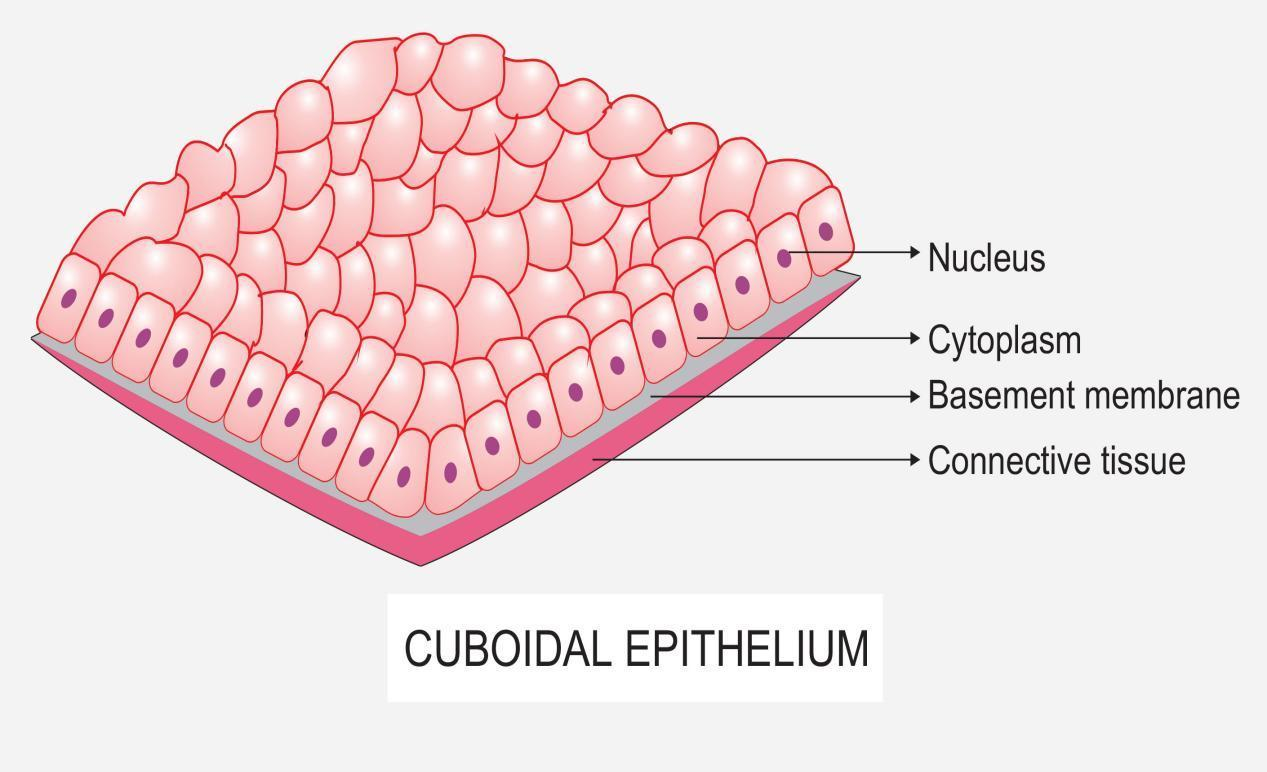
Each cell has a nucleus. This tissue consists of cubical cells. With large, rounded, centrally located nuclei, all the cells of this epithelium are directly attached to the basement membrane. A columnar epithelial cell looks like a column or a tall rectangle. This epithelial type is found in the small collecting ducts of the kidneys,.

Simple Epithelium Tissue
Small cubes in cross section 3) columnar : Identification of stratified squamous epithelium. Each cell has a nucleus. Web simple cuboidal epithelium consists of a single layer cells that are as tall as they are wide. Secretory ducts of small glands, kidney tubules. A squamous epithelial cell looks flat under a microscope. Web introduction to.
Simple cuboidal epithelium medical images for power point
The important functions of the simple cuboidal epithelium are secretion and absorption. Web the first pages illustrate introductory concepts for those new to microscopy as well as definitions of commonly used histology terms. A squamous epithelial cell looks flat under a microscope. They are sketches from selected slides used in class from the. Simple cuboidal.

Simple Cuboidal sldie Labeled Histology Epithelial Tissues
A squamous epithelial cell looks flat under a microscope. Thin and flat 2) cuboidal : The basement membrane is a thin but strong, acellular layer which lies between the epithelium and the adjacent connective tissue. Blood and lymphatic vessels, air sacs of lungs, lining of the heart. Web simple cuboidal epithelia are observed in the.

Simple cuboidal epithelium, illustration Stock Image C052/3697
A columnar epithelial cell looks like a column or a tall rectangle. Web function and location of simple epithelium. Thin and flat 2) cuboidal : The drawings of histology images were originally designed to complement the histology component of the first year medical course run prior to 2004. One layer of cells 2) stratified :.
Simple Cuboidal Drawing They are sketches from selected slides used in class from the. Want to create or adapt books like this? A columnar epithelial cell looks like a column or a tall rectangle. Web simple cuboidal epithelium consists of a single layer cells that are as tall as they are wide. Web simple cuboidal epithelium:
They Are Sketches From Selected Slides Used In Class From The.
Every cell attaches to the basement membrane. A cuboidal epithelial cell looks close to a square. Web simple cuboidal epithelium: The important functions of the simple cuboidal epithelium are secretion and absorption.
Want To Create Or Adapt Books Like This?
First, we will draw the simple cuboidal epithelium lining a kidney tubule. Each cell has a nucleus. One layer of cells 2) stratified : Web the first pages illustrate introductory concepts for those new to microscopy as well as definitions of commonly used histology terms.
Web 77K Views Simple Cuboidal Epithelium Location Simple Cuboidal Epithelium Is Located Deep Within The Body Where It Lines Secretory Ducts And Tubules In Several Organs.
Web introduction to simple cuboidal epithelium. Thin and flat 2) cuboidal : This epithelial type is found in the small collecting ducts of the kidneys, pancreas, and salivary glands. Secretory ducts of small glands, kidney tubules.
Web Three Main Shapes Of Cells At The Apical/Free Surface 1) Squamous :
Identification of simple columnar epithelium. Web function and location of simple epithelium. A squamous epithelial cell looks flat under a microscope. Simple cuboidal cells are also characterized by a single, large, round (spherical.