
Simple Cuboidal Epithelium Drawing
Simple Cuboidal Epithelium Drawing - Functions by lining the surface of various ducts of various glands and organs, simple cuboidal cells are able to provide a layer of protection from abrasion, foreign particles, invading bacteria and excessive water loss (due to its selective permeability) to the underlying tissue. Secrets lubricating substance, allows diffusion and filtration. You will notice many tubules in this view. Simple cuboidal epithelia are observed in the lining of the kidney tubules and in the ducts of glands. Diagram of simple cuboidal epithelium.
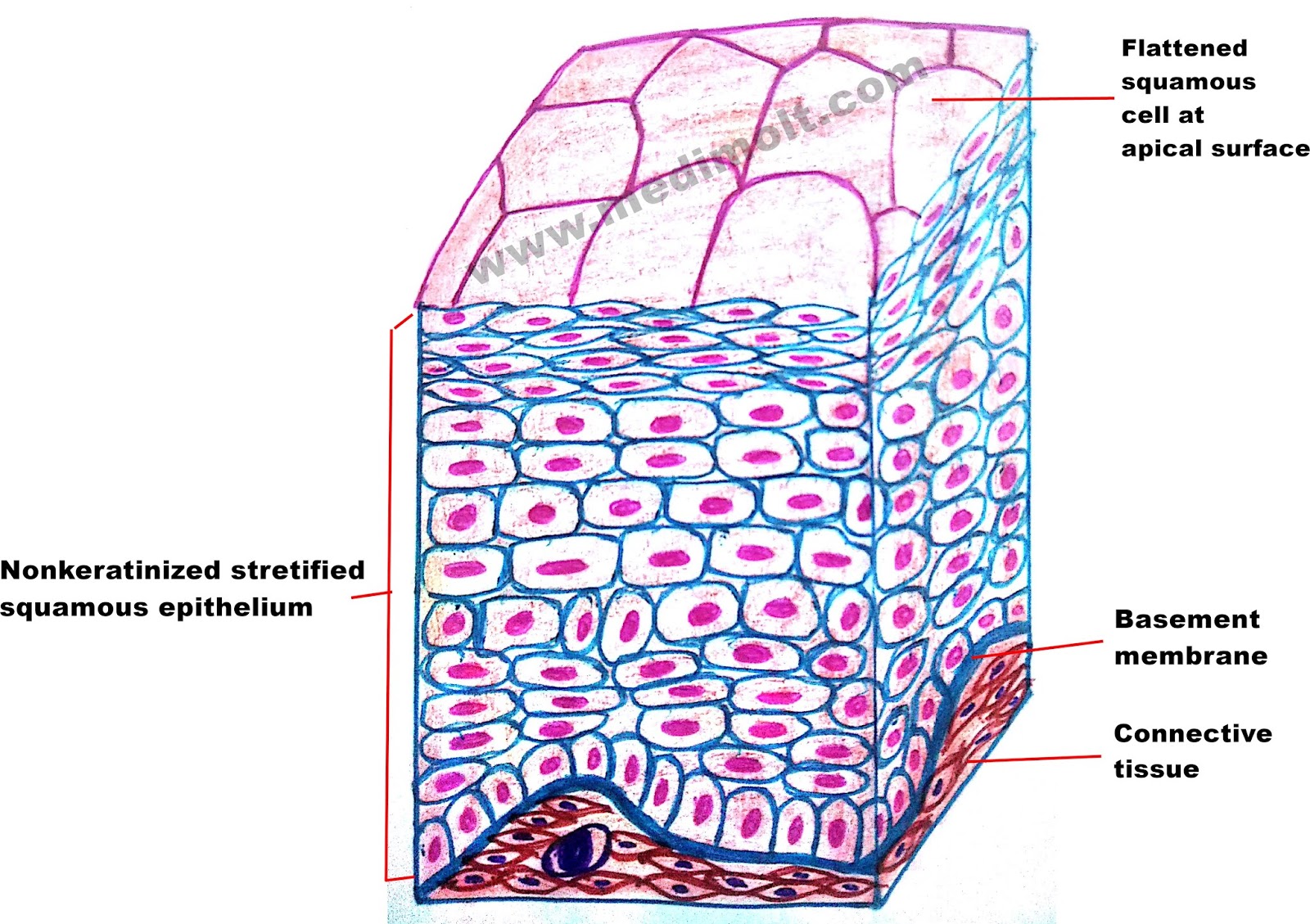
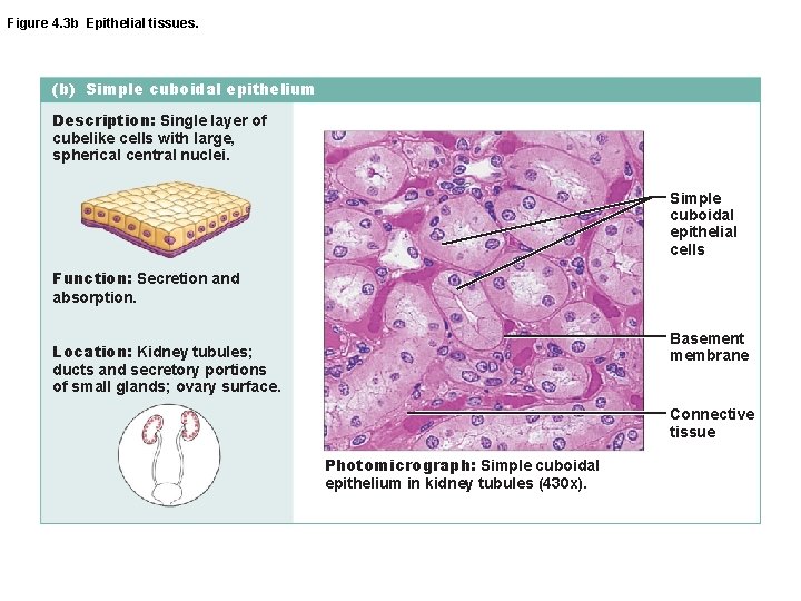
Sweat glands, mammary glands, developing ovarian follicles. Web simple cuboidal epithelium definition. Web drawing histological diagram of simple cuboidal epithelia. These cuboidal cells have large, spherical and central nuclei. Blood and lymphatic vessels, air sacs of lungs, lining of the heart. This tissue consists of cubical cells. With large, rounded, centrally located nuclei, all the cells of this epithelium are directly attached to the basement membrane.

Simple cuboidal epithelium Diagram Quizlet
Read more about simple cuboidal thyroid gland 40x; Web simple cuboidal epithelia are like cubes and usually there is as much cytoplasm over the nucleus as there is under it. You can see the single layer of cube shaped cells of each tubule. This tissue consists of cubical cells. Simple cuboidal epithelium (400x) kidney cortex.
Simple Cuboidal Epithelium Location
The above section shows simple cuboidal epithelia in the thyroid gland. Desai here the tubules are shown in longitudinal section. Explanation on epithelia while drawing. Web simple cuboidal epithelium (two cells indicated by black box) make up the tubules in the kidneys (shown in cross section and indicated by the black oval). Simple cuboidal cells.

Simple cuboidal epithelium Diagram Quizlet
Web welcome to diya's art tutorial youtube channel today in this video i'm showing how to draw cuboidal epithelial tissue (step by step) |. Web drawing histological diagram of simple cuboidal epithelia. Secrets lubricating substance, allows diffusion and filtration. They are mostly derived to suit the function of the particular organs better. Explanation on epithelia.

What Is The Main Function Of Simple Cuboidal Epithelium? Mastery Wiki
Thyroid gland, 40x objective 400x total magnification, simple cuboidal. Drawn by using h & e pencils. These cuboidal cells have large, spherical and central nuclei. Simple cuboidal epithelia are observed in the lining of the kidney tubules and in the ducts of glands. Web simple cuboidal epithelia are observed in the lining of the kidney.

Simple Cuboidal Epithelium Histology Pinterest Anatomy Images and
Web introduction to simple cuboidal epithelium. They are mostly derived to suit the function of the particular organs better. A cuboidal epithelial cell looks close to a square. Secretory ducts of small glands, kidney tubules. Web simple cuboidal epithelia are observed in the lining of the kidney tubules and in the ducts of glands. Web.

Simple Cuboidal Epithelium Kit Ng, Ph.D.
Web there are three basic shapes used to classify epithelial cells. The above section shows simple cuboidal epithelia in the thyroid gland. Web type of cuboidal epithelium. Secrets lubricating substance, allows diffusion and filtration. These cell lies on a basement. Diffusion and absorption are the processes by which epithelial cells move substances out of the.

How to draw stratified cuboidal epithelium easy way YouTube
Web simple cuboidal epithelium functions include diffusion, absorption, secretion, and protection. Simple cuboidal epithelium (400x) kidney cortex These epithelia are involved in the secretion and absorptions of molecules requiring active transport. Web histology easy diagrams 2021.simple cuboidal epithelium dr khushboo mogra 1.18k subscribers subscribe 14 share 533 views 1 year ago Blood and lymphatic vessels,.
Simple Cuboidal Epithelium Labeled Cell Membrane Drawo
Web simple cuboidal epithelia are observed in the lining of the kidney tubules and in the ducts of glands. Web type of cuboidal epithelium. Web simple cuboidal epithelium definition. Web since you are trying to find a simple cuboidal epithelium, look for rows or rings of round dark dots (the nuclei of the cuboidal epithelial.

Simple Epithelium Tissue
Lining the kidney tubules, the ovary, thyroid. You can see the single layer of cube shaped cells of each tubule. Explanation on epithelia while drawing. Secretory ducts of small glands, kidney tubules. They are mostly derived to suit the function of the particular organs better. Drawn by using h & e pencils. Simple cuboidal cells.
Simple cuboidal epithelium medical images for power point
Read more about simple cuboidal thyroid gland 40x; With large, rounded, centrally located nuclei, all the cells of this epithelium are directly attached to the basement membrane. Simple cuboidal cells are also characterized by a single, large, round (spherical. The above section shows simple cuboidal epithelia in the thyroid gland. Most of the cells in.
Simple Cuboidal Epithelium Drawing A squamous epithelial cell looks flat under a microscope. Lining the kidney tubules, the ovary, thyroid. Web simple cuboidal epithelia are observed in the lining of the kidney tubules and in the ducts of glands. Diffusion and absorption are the processes by which epithelial cells move substances out of the fluid. Read more about simple cuboidal thyroid gland 40x;
Web Histology Easy Diagrams 2021.Simple Cuboidal Epithelium Dr Khushboo Mogra 1.18K Subscribers Subscribe 14 Share 533 Views 1 Year Ago
Blood and lymphatic vessels, air sacs of lungs, lining of the heart. Drawn by using h & e pencils. Secrets lubricating substance, allows diffusion and filtration. Web simple cuboidal epithelium definition.
Simple Cuboidal Epithelium (400X) Kidney Cortex
Most of the cells in this image are simple cuboidal epithelial cells. Diffusion and absorption are the processes by which epithelial cells move substances out of the fluid. Thyroid gland, 40x objective 400x total magnification, simple cuboidal. Secretory ducts of small glands, kidney tubules.
Web Simple Cuboidal Epithelia Are Observed In The Lining Of The Kidney Tubules And In The Ducts Of Glands.
Web simple cuboidal epithelia are observed in the lining of the kidney tubules and in the ducts of glands. Explanation on epithelia while drawing. Web drawing histological diagram of simple cuboidal epithelia. Simple cuboidal cells are also characterized by a single, large, round (spherical.
Web Since You Are Trying To Find A Simple Cuboidal Epithelium, Look For Rows Or Rings Of Round Dark Dots (The Nuclei Of The Cuboidal Epithelial Cells).
Web simple cuboidal epithelium (two cells indicated by black box) make up the tubules in the kidneys (shown in cross section and indicated by the black oval). Sweat glands, mammary glands, developing ovarian follicles. They are mostly derived to suit the function of the particular organs better. Each cell have centrally located round nucleus.