
Drawing Of Paramecium
Drawing Of Paramecium - Web this will also help you to draw the structure and diagram of paramecium. Flexible, thin, elastic membrane consisting of outer plasma membrane and an inner membrane called epiplasm, but lacking a cell wall. Web download 75 paramecium diagram stock illustrations, vectors & clipart for free or amazingly low rates! Higher magnification of the metachronal. Pellicle protects the cell from the outside environment.
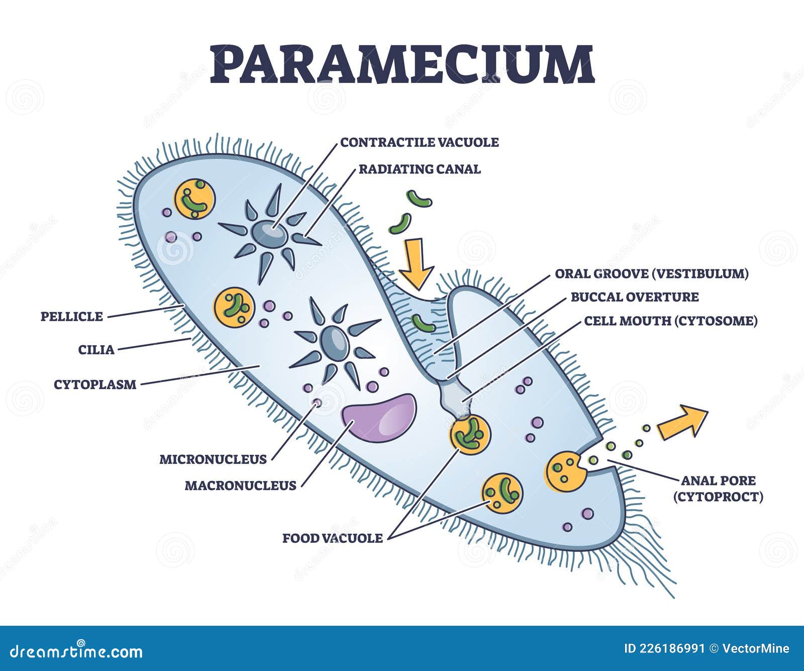
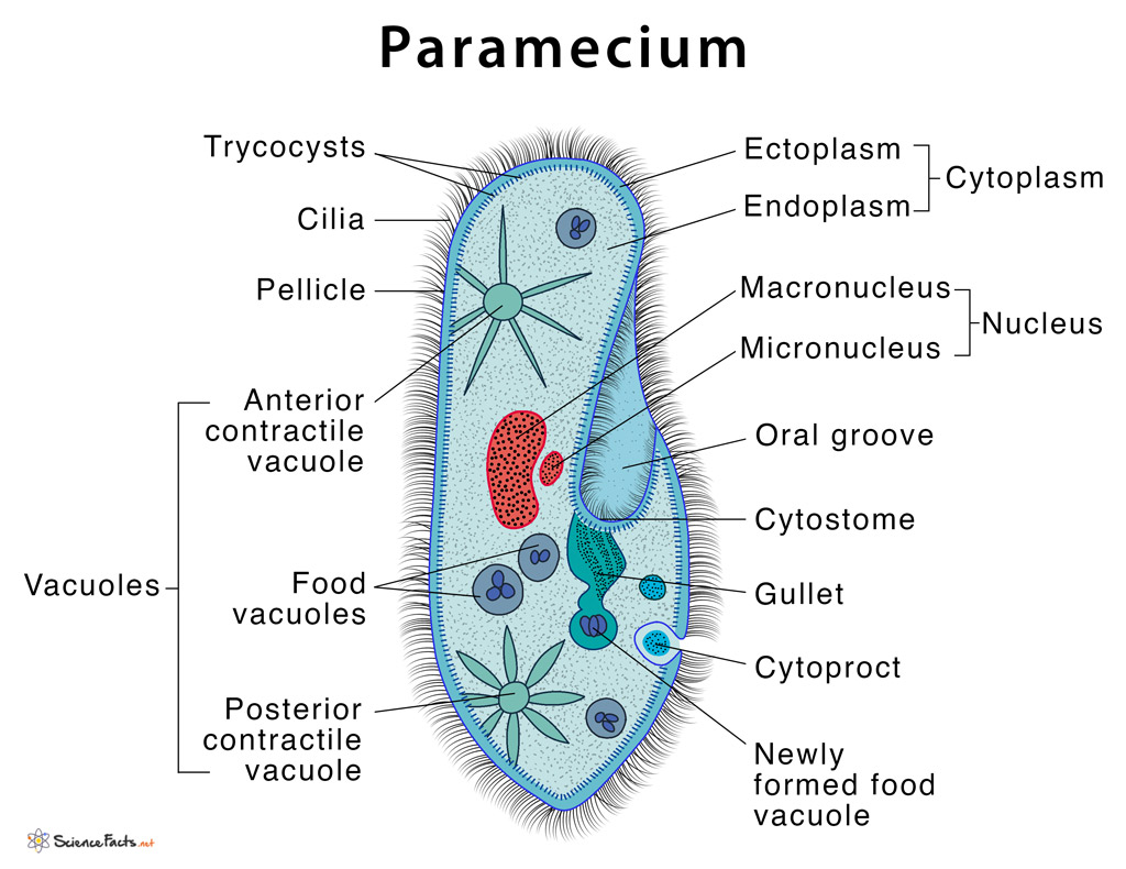
Please use the first image to make a sketch (drawing) of paramecium on a piece of paper. Web this will also help you to draw the structure and diagram of paramecium. How to draw paramecium step by step | easy paramecium diagramhello friends in this video i will tell you about how to draw labelled diagram of paramec. Oral groove cytostome, cytopharynx, and food. Cv contractile vacuoles, fv food vacuoles, manu macronucleus, mino micronucleus, pe peristome, tr trichocysts and ve vestibulum. Paramecium is a unicellular organism with a shape resembling the sole of a shoe. Web this is lined with inconspicuous cilia which beat continuously, drawing food into the cell.
Paramecium Microscopic Closeup Structure with Anatomical Outline
Paramecium are primarily heterotrophic, feeding on bacteria and other small organisms. It ranges from 50 to 300um in size. Higher magnification of the metachronal. Web appearance paramecia cells are elongated in appearance, and based on this shape were divided into two groups: Surprisingly, paramecium is visible to the naked eye and has an elongated slipper.

How To Draw Paramecium Diagram YouTube
It is the pencil diagram of paramecium for class 10, 11 and 12. Web panel 1 (a,b): Please use the first image to make a sketch (drawing) of paramecium on a piece of paper. Paramecium is a ciliate protozoan and moves by beating of cilia, called ciliary. Cv contractile vacuoles, fv food vacuoles, manu macronucleus,.

DRAW IT NEAT How to draw Paramecium
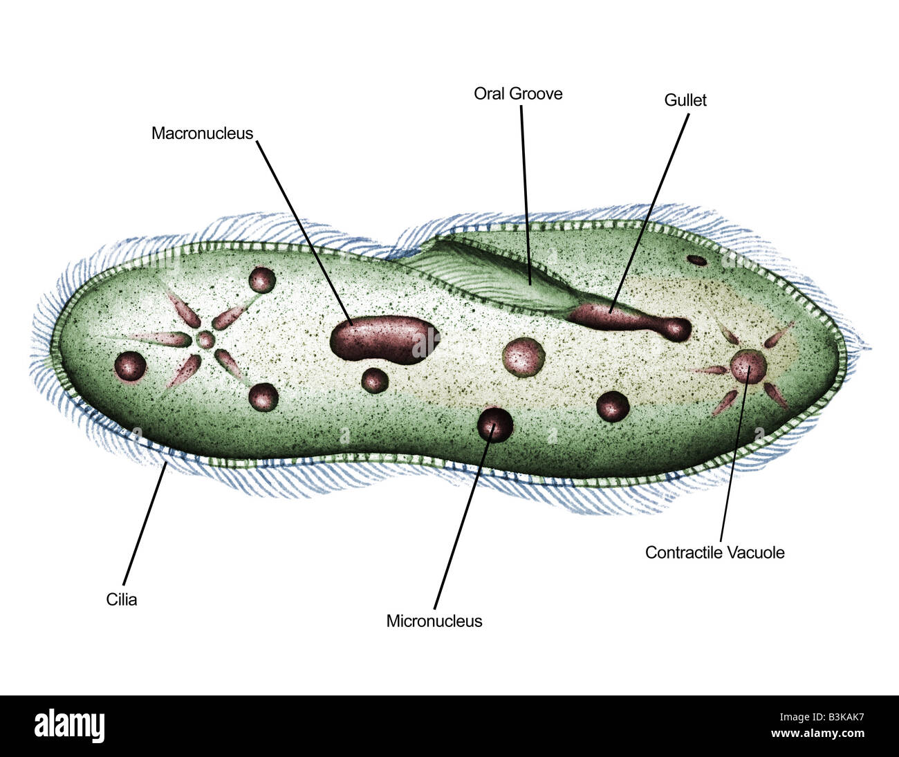
Web panel 1 (a,b): Use the second image as a guide to label the structures on your sketch. Web this is lined with inconspicuous cilia which beat continuously, drawing food into the cell. Drawing of paramecium illustrating light microscopic features: Take a picture of your sketch and embed it here. Web this video helps you.
Paramecium Definition, Structure, Characteristics and Diagram
How does a paramecium eat? Then determine the size utilizing a scale bar. The common species of paramecium include: New users enjoy 60% off. Web 4 30 views 8 months ago easy science drawing how to draw a labelled diagram of paramecium. Surprisingly, paramecium is visible to the naked eye and has an elongated slipper.

Paramecium Diagram, Homeschool science, Print
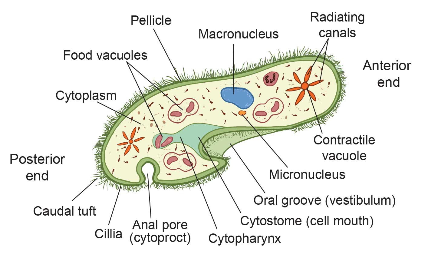
Oral groove cytostome, cytopharynx, and food. Web the basic anatomy of paramecium shows the following distinct and specialized structures in their cell: Paramecia live mainly by heterotrophy, feeding on bacteria and other small organisms. They are ciliated protozoan and come under phylum ciliophora. Web the anatomy of paramecium paramecium wears a soft armor, called pellicle.

Paramecium Diagrams to Print 101 Diagrams
Higher magnification of the metachronal. The term paramecium is also used to refer to individual organisms in a paramecium species. Web #paramecium #howtodraw #biologythis is a diagram of the paramecium. Unlike amoeba, paramecium has a distinct and permanent shape and certain areas of cytoplasm, (cell organelles), are specialised to carry out specific functions. Then determine.

DRAW IT NEAT How to draw Paramecium
Flexible, thin, elastic membrane consisting of outer plasma membrane and an inner membrane called epiplasm, but lacking a cell wall. Paramecia live mainly by heterotrophy, feeding on bacteria and other small organisms. Paramecium is a ciliate protozoan. Pellicle protects the cell from the outside environment. Web #paramecium #howtodraw #biologythis is a diagram of the paramecium..
ILLUSTRATED DIAGRAM OF PARAMECIUM (PARAMECIUM SP.) 1000X Stock Photo
Web this will also help you to draw the structure and diagram of paramecium. Paramecia live mainly by heterotrophy, feeding on bacteria and other small organisms. Pellicle protects the cell from the outside environment. Fresh water, free living, omnipresent and is found in stagnant water. Cv contractile vacuoles, fv food vacuoles, manu macronucleus, mino micronucleus,.

Structure Of A Paramecium stock vector art 499581981 iStock
Web in this video i have shown the simplest way of drawing paramecium. Then determine the size utilizing a scale bar. Web 4 30 views 8 months ago easy science drawing how to draw a labelled diagram of paramecium. Its size ranges from 170 to 290um or up to 300 to 350um. Their basic shape.
The Structure of Paramecium Cell Rs' Science
Higher magnification of the metachronal. Cadatum is a microscopic, unicellular protozoan. Flexible, thin, elastic membrane consisting of outer plasma membrane and an inner membrane called epiplasm, but lacking a cell wall. Its size ranges from 170 to 290um or up to 300 to 350um. Take a picture of your sketch and embed it here. New.
Drawing Of Paramecium Please use the first image to make a sketch (drawing) of paramecium on a piece of paper. Web panel 1 (a,b): Paramecium is a ciliate protozoan. Surprisingly, paramecium is visible to the naked eye and has an elongated slipper like shape, that’s the. Their basic shape is an elongated oval with rounded or pointed ends, such as in p.
Drawing Paramecium And Labeling Is Shown In This Diagram.
Web this will also help you to draw the structure and diagram of paramecium. Its size ranges from 170 to 290um or up to 300 to 350um. Web this is lined with inconspicuous cilia which beat continuously, drawing food inside the cell. They are ciliated protozoan and come under phylum ciliophora.
Drawing Of Paramecium Illustrating Light Microscopic Features:
Light microscopic appearance of paramecium caudatum. Unlike amoeba, paramecium has a distinct and permanent shape and certain areas of cytoplasm, (cell organelles), are specialised to carry out specific functions. Then determine the size utilizing a scale bar. Higher magnification of the metachronal.
A Few Species Are Mixotrophs, Deriving Some Nutrients From Endosymbiotic Algae Carried In The Cytoplasm Of The Cell.
Paramecium is unicellular and eukaryotic, so they are kept in the kingdom protista. Paramecium diagram is given below. The term paramecium is also used to refer to individual organisms in a paramecium species. It ranges from 50 to 300um in size.
Structure Of Paramecium Diagram Paramecium Locomotion.
Web the anatomy of paramecium paramecium wears a soft armor, called pellicle paramecium’s skin is covered by many tiny hairs, called cilia the microscopic view of cilia the structure of pellicle and cilia see how cilia do the wave how fast can a paramecium move? Web this video helps you to draw science diagrams with great ease and clarity. Take a picture of your sketch and embed it here. Cv contractile vacuoles, fv food vacuoles, manu macronucleus, mino micronucleus, pe peristome, tr trichocysts and ve vestibulum.